Download
1 / 66
660 likes | 875 Views
Tissue classified according to regeneration potential Labile - continues to regenerate throughout life (“continuous replicators”) Note: any type of tissue regeneration requires basement membrane Examples: Skin, GI tract, vagina, endometrium , bone marrow, lymph,

E N D
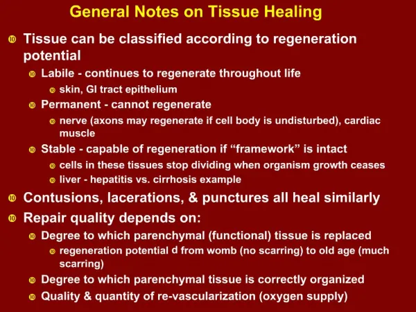
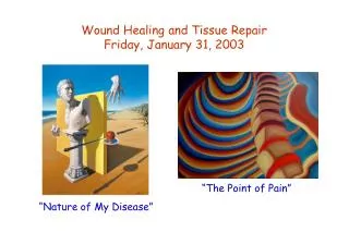
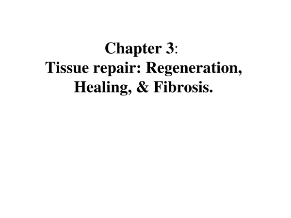
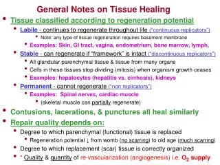
Tissue classified according to regeneration potential • Labile - continues to regenerate throughout life (“continuous replicators”) • Note: any type of tissue regeneration requires basement membrane • Examples: Skin, GI tract, vagina, endometrium, bone marrow, lymph, • Stable - can regenerate if “framework” is intact (“discontinuous replicators”) • All glandular parenchymal tissue & tissue from many organs • Cells in these tissues stop dividing (mitosis) when organism growth ceases • Examples: hepatocytes (hepatitis vs. cirrhosis), kidneys • Permanent - cannot regenerate (“non replicators”) • Examples: Spinal nerves, cardiac muscle • (skeletal muscle can partially regenerate) • Contusions, lacerations, & punctures all heal similarly • Repair quality depends on: • Degree to which parenchymal (functional) tissue is replaced • Regeneration potential ↓from womb (no scarring) to old age (much scarring) • Degree to which replacement (scar) tissue is correctly organized • * Quality & quantity of re-vascularization(angiogenesis)i.e. O2 supply General Notes on Tissue Healing
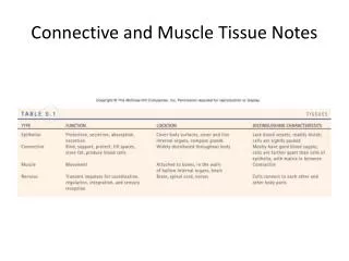
Skin Stratum corneum (horny layer) (keritinized cells) Stratum granulosum (granular layer) Epidermis Stratum germinativum (basil layer) Dermal papillae (form ridges) Elastic Fibers Collegen Fibers Reticular Fibers Fibrocytes Blood Vessels Sensory organs Sweat Glands Dermis (Dense Irregular Tissue) Adipose tissue Areolar fibers Hypodermis (Subcutaneous) Surface Area: 1.8 to 2.0 square meters ↑7 X from birth to adult Thickness: .5 to 3.0 mm average 1 to 2 mm
Skin Micrograph Stratum corneum (horny layer) (keritinized cells) Stratum granulosum (granular layer) Stratum germinativum (basil layer) Dermal papillae (form ridges) Elastic Fibers Collegen Fibers Reticular Fibers Fibrocytes Blood Vessels Sensory organs Sweat Glands Adipose tissue Areolar fibers
Skin • Epidermis - avascular but well innervated - .3mm to 1mm thick • Regenerated in normal healing • Stratified squamous epithelium • Deeper cells (stratum germinativum) are the only “living” cells • Deeper cells become increasingly keratinized as they are pushed to surface • Epidermal mitosis is stimulated by the loss of horny layer cells • More active at night • Dermis - .5mm to 3 mm thick • Regenerated in normal healing • Dermal papillae helps hold epidermis in place (ridges - finger prints) • Dermis has smooth muscle tissue (pilo-erector) bundled with hair follicles • Prominent examples (scrotum, areola) • Langer’s lines - lines formed at right angles to direction of natural stretch • Caused by gravity & activity • Incisions along these lines tend to form less scar tissue • Substantial plastic surgery implications • Hypodermis - Subcutaneous tissue • Density & cellular arrangement of this tissue determines skin “mobility” • Composed mostly of fat cells and areolar tissue
Healing of Lacerations, Abrasions & Punctures • 1. Inflammatory Phase: inflammation, bleeding / clotting (0 - 48 hours) • Ruptured cell membranes →reflexes, thromboxane & prostaglandins cause temporary vasoconstriction • Thromboplastin & Hageman factor & platelets produce platelet aggregation: • Activation of compliment system • Attraction of neutrophils & inflammatory mediators (chemotaxis) • Arachidonic acid cascade → ↑prostaglandins, thromboxane, & leukotriene • Mast cells & basophils release histamine → ↑vascular permeability • Blood touches collagen→platelet activation • Plug formation/aggregation, coagulation system activation, cytokine & growth factor release • Release of serotonin, histamine, prostaglandins →↑ vascular permeability • PDGF & cytokines begins attracting fibroblasts • Cytokines orchestrates inflammation and the cellular activity involved in healing • Activation of kalikrein→ ↑bradykinin→PAIN& vasodilation • Fibrin & Fibronectin form protective scabhttp://www.emedicine.com/ent/TOPIC13.HTM • Surface Coagulation completed - Granulation tissue forms • Neutrophils begins to arrive within an hour after wounding • Macrophages release angiogenic growth factors that stimulate re-vascularization • GRANULATION TISSUE:capillary buds, fibroblasts, macrophages • Scar forming tissue
Healing of Lacerations, Abrasions & Punctures • 2. Fibroplastic (proliferatory) phase - begins during inflammatory phase !!! • Tissue hypoxia + macrophage GF →fragile capillary “extensions” or “buds” • Usually happens within 24 to 72 hours after injury • Termed “angiogenesis” • Buds proliferate & grow new circuits & also connect with existing capillaries • Capillary – Venule loops are formed • Fibroblasts→fibronectin and additional extracellular matrix components • Proteoglycans, collagen fibers, reticular fibers, elastic fibers, glycoproteins • Vitamin C & oxygen needed for collagen synthesis • In extracellular matrix (ECM), hyaluronic acid combines with fibronectin • Fibronectin + hyaluronic acid →“scaffold” or “framework” for cell migration • Fibronectin - adhesive glycoprotein located in blood & cell membranes • Initial wound tensile strength (day 4 or 5) provided by: • Fibronectin & cross-linking of fibronectin & collagen • Keratinocytes regenerate epithelium and it proliferates beneath the clot • Wound contraction occurs • Myofibroblasts located in wound margins have high actin content • Myofibroblasts move toward the center of the wound & contract • Ends of damaged tissue are pulled closer together
Healing of Lacerations, Abrasions & Punctures • 3. Maturation phase - mostly complete in 3 weeks to 6 months • Clot lysis (early in the phase) • Breakdown & re-synthesis of collagen fibers in direction of tensile forces • May continue for years • Elevated pink scar eventually replaced soft flat white scar • Embryonic Type III collagen (inferior) replaced by normal Type I collagen
Cell Recruitment in the Wound Coagulation Inflammation Fibroblastic Proliferation Remodelling Neutrophils Platelets Macrophages Fibroblasts Relative Number of Cells Lymphocytes 16 4 14 2 8 12 10 0 6 Days Post-Wounding
Wound Healing: Inflammatory Phase Clot Keratinocytes produce New Epithelial Tissue Granulocytes (neutrophils macrophages) Early Granulation Tissue Capillary Bud Formation (Angiogenesis)
Wound Healing: Fibroblastic Phase Collagen Bundles in Dermis Capillary Buds Finally Form Capillary-Venule Loop Mitosis of Epithelial Tissue Fibroblasts (migrating along “scaffold”) New Collagen Fibers Fibronectin ECM “Scaffold” Granulocytes
Wound Healing: Early Maturation Phase Epithelial Migration Complete New Collagen Fibers (Type III Collagen - Scar Tissue) Note tissue disorganization
Capillary Angiogenesis • Excessive angiogenesis occurs in diseases such as: • Cancer • Diabetic blindness • Age-related macular degeneration • Rheumatoid arthritis and • Psoriasis • Insufficient angiogenesis occurs in conditions such as: • Coronary artery disease • Stroke • Delayed wound healing ↓O2tension in granulation tissue • Dead & injured tissue along with macrophages release growth factors that bind to receptors on capillary endothelium • Proteases released from activated endothelial cells degrade basement membrane • Endothelium proliferates into granulation tissue • The enzyme MMP dissolves tissue in front of the sprouting vessels • Integrins (proteins) “grapple” the granulation tissue and pull the capillary buds forward • Sprouting endothelial buds roll up and form a blood vessel tube which is covered with fibronectin and proteogylcans • Tube connect to form loops with other neo-vascular tissue and existing capillaries Note: control of angiogenesis is through the balance of angiogenic growth factors and angiogenic inhibitors. Normally, the balance favors the production of inhibitors http://www.angio.org/understanding/understanding.html
Healing by First & Second Intention • Healing by first intention - wound edges brought together by sutures • ↓loss of parenchymal tissue → ↓amount of scar tissue • Healing occurs faster • Less chance of infection • Healing by second intention- wound not closed / unable to be closed • Examples: decubitus ulcers (bed sores), burns • Significant loss of parenchymal tissue → ↑amount of scar tissue • Healing occurs slower • Greater chance of infection
Wound Healing First Intention (sutures) Second Intention (no sutures) Larger Tissue Defect More Scar Tissue
Things that Inhibit Healing • Ischemia (lack of oxygen to the wound) • Poor blood supply • Dry Wound Environment • Cover wounds & keep them moist with antibiotic ointment • Infection • Make sure wound is thoroughly cleaned before bandaging • Foreign Bodies or Material Left in Wound • Change bandage often and look for foreign material • NSAIDAnti-inflammatory Medications • Nutritional deficiency
Granulation Tissue Becomes Scar Tissue • Scar tissue is not as vascularized, flexible, elastic, or strong as original tissue • Scar tissue formed in a muscular organ may inhibit function • Examples: heart, bladder • Scar tissue may form Adhesions which connect adjacent organs • Adhesions:inflammatory fibrous bands connecting serous surfaces • Most often found in pleural cavity and peritoneal cavities • May cause loss of function • Scar tissue may formContractures • Contractures: Fibrotic tissue laid down in connective tissue, skin, fascia, muscle, or joint capsule that prevents normal movement • May form within a joint→loss of mobility & range of motion • May form in skin or muscle fascia →loss of mobility & elasticity • Stretch Marks(striaedistensae) – results from tearing of the dermis • Fibroblasts cannot secrete enough fibers to keep rapidly growing skin taut • May have a genetic influences • Usually appears where large amounts of fat are stored • Abdomen, breasts, thighs, hips, butt, upper arms, under arms • Approximately 90% of pregnant women, 70% of adolescent females, and 40% of adolescent males have stretch marks. http://www.webmd.com/baby/features/stretch-marks-getting-under-your-skin?src=RSS_PUBLIC
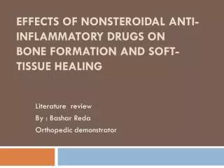
Classification of Muscle Injury • Muscle Strain - tension exceeds the weakest structural element • Injury usually located 0.1 to 3 mm from the muscle-tendon junction • Caused by: sudden over-stretch or contraction and limb deceleration • Failure of GTO may play a significant role in muscle strain • Insufficient warm-up may contribute to muscle strain • Contributory causes: corticosteroid injections & previous injury • First degree (mild) • Minimal structural damage and minimal hemorrhage • Second degree (moderate) • Partial tissue tear, significant loss of function, significant hemorrhage • Third degree (severe) • Complete tear, total loss of function, extensive hemorrhage, possible muscle (myofibrilllar) retraction • http://www.emedicinehealth.com/muscle_strain/article_em.htm • http://www.sportsinjuryclinic.net/cybertherapist/back/hamstrings/hamstringstrain.htm
Classification of Muscle Injury • Muscle Contusion: blow to muscle → fiber tearing→hematoma • Severe contusions sometimes difficult to distinguish from complete tear • Intermuscular hematoma - bleeding between muscle fascia • Characterized by early migration of ecchymosis to distal part of limb • Heals more quickly than intramuscular hematomas • Intramuscular hematoma - bleeding within a fascia enclosed muscle bundle • Hemorrhage is more confined and localized • Inflammatory response is exaggerated • ↑risk of myositis ossificans, scarring, & compartment syndrome • Compartment syndrome: hemorrhage → ↑pressure in muscle unit • ↑pressure → ↓blood flow →ischemia→necrosis→gangrene • Most often result from severe contusions • Can be caused by excessive exercise induced muscle damage • Causes severe pain, palpable tightness, “shiny” skin appearance • Fascia release (surgery) is done within 12 hrs to minimize damage
General Steps in the Healing of Muscle Trauma Stage Pathology - Healing Treatment Implications Inflammatory- Cell disruption→hemorrhage & edema formation - RICE (days 0 - 2) - Myofibrillar retraction + possible nerve axon damage - Immobilization & Protection - PMN and MN cell invasion (inflammation) - NSAID drugs - Phagocytosis Fibroplastic-↑rate of collagen synthesis by fibroblasts - Partial ROM exercises Proliferation- Muscle regeneration from 2 types of “Satellite” cells- Gentle resistance work (days 3 - 6) (Satellite cells are “stem cells” activated by injury)-Directionalize collagen & muscle - Neovascularization - Protect neovascularization - Pulsed ultrasound Fibroplastic- Muscle fiber & satellite cell fusing & bridging begins -↑resistance work Proliferation- Tensile strength approximately 50% of normal - Progress to full ROM exercises (days 7 - 14) - Contraction still inhibited by edema & pain - Ultrasound & heat modalities - Tendency to try to “return to action” →↑chance of re-injury Remodeling-↑maturation of collagen → ↑tensile strength - Progression of activity Maturation- Replacement of initial or inferior tissues -↑intensity & duration ( days 15 - 60) - Permanent loss of tissue tensile strength: 7%
MRI of Adductor Longus Muscle Strain- Note ↓ signal intensity (very dark) around old scar & cortical bone of femur- Note ↑signal intensity around the strain indicating fluid buildup L R
MRI of Muscle Strain MRI of adductor longus strain (sagital) Note retraction of muscle (red arrows) Normal contralateral muscle (green arrow) MRI of biceps femoris (hamstring) strain Note ↑signal intensity →edema (green arrow) Myofibrillar Retraction
CT-scan of Intramuscular Hematoma CT Scan of intramuscular hematoma from a direct blow to the quadriceps Note the decreased signal intensity (arrow) indicating fluid buildup This particular person applied heat to the injury, exacerbating it An emergency fasciotomy had to be performed
MRI of Myositis Ossificans Myositis Ossificans in Vastus Lateralis Femur
Properties of Ligamentous and Tendinous Tissues 2x cross sectional area steeper slope →greater tissue “stiffness” Deformation Force (Stress or Load) x cross sectional area Change in Tissue Length (Strain or % Deformation) x length Deformation Force (Stress or Load) 2x length Change in Tissue Length (Strain or % Deformation)
Ligament Injury • Ligament - fibrous dense connective tissue - binds bones • Injuries to these structures are associated with the future development of OA • Ligaments have subunits that tighten or loosen depending on joint position • Ligaments are not densely innervated or densely vascularized • Do contain some blood vessels and nerves in outer covering (epiligament) • Do contain proprioceptors • Do transmits pain signals via type C fibers • In bone-ligament-bone structures, ligament is the weakest link • Weakest near ligament insertion (adolescent & osteoporotic exceptions) • Ligaments are not readily weakened by inactivity (takes many weeks) • Ligaments show only a 10% - 20% ↑in tensile strength with exercise training • Surgical repair not done unless ends are significantly far apart • Length of repair scar does not affect tensile strength • Long repair scar → ↓joint stability &↑joint laxity • ACL tears most often result in ends unopposed →surgery required • Surgical repair restores only about 80% - 90% of original tensile strength
Functional Sub-units of the Lateral Collateral Ligament - Left Knee
Ligament Sprain • In most cases, more than 1 ligament share loads around a joint • Most sprains involve more than one ligament - example: ankle • Most common sprain: ankle inversion accompanied by plantar flexion • Primary ligaments: anterior talofibular ligament and calcaneofibular ligament • If sprain is severe, “backup” structures may sometimes be involved • Backup structures: posterior talofibular ligament & peroneal tendon • Repeat injuries not only tear healed areas but backup structures as well • Prevention of re-injury is of critical importance • Most common knee sprain: valgus force to knee → medial collateral tear • Backup structure: anterior cruciate (cruciates’ blood supply inferior to collaterals’) • Joint instability in knee sprain likely to be evident only in injury position • Ligament sprain classifications • Grade I - Slight incomplete tear - no notable joint instability • Grade II - Moderate / severe incomplete tear - some joint instability • One ligament may be completely torn • Grade III - Complete tearing of 1 or more ligaments - obvious instability • Surgery usually required http://catalog.nucleusinc.com/displaymonograph.php?MID=7
High Ankle Sprain A "high ankle sprain”,also known as “syndesmotic sprain”, is an inury to the syndesmotic and / or “Interosseous ligaments” that joins the tibia and fibula bones of the lower leg. The distinction exists because a "high" ankle sprain occurs above the ankle joint and is more severe than a sprain of the joint ligaments. A high ankle sprain results from an outward twisting of the ankle
Ligament Healing Stage Pathology - Healing Treatment Implications InflammatoryIntra-articular injury (ACL & PCL)- RICE (Protect & Immobilize <48 hrs) (days 0 - 4) -↑intra-articular pressure & hemarthrosis- Immobilize (→ ↓osteoarthritis) Extra-articular injury (LCL & MCL)- NSAID drugs - Subcutaneous hematoma - Light passive ROM exercise machines - Exercises that “cross” the joint (straight leg raises for ACL injury) - Weight bearing exercise & mobilization begun as soon as possible Fibroplastic-Fibroblasts & angiogenic cells →scar matrix - Progress to full active ROM exercise Proliferation- Phagocytes remove damaged ligament debris - Resistance exercise (day 4 - weeks) - “Decent” tensile strength within 3 weeks -↑intensity of all types of exercises - ACL & PCL heal using fibrocartilagenous cells - Biomechanical evals began at 3 wks Remodeling-↑density of scar matrix - Progression of activity Maturation- Replacement of initial or inferior collagen tissues (↑intensity & duration) (weeks to years) -↑strength of molecular bonds of scar matrix - Near maximum strength reach within 1 year ** but not back to 100% of original Healed Ligament never attains pre-injury tensile strength due to: ↓Number of hydroxypyridiniumcross linkages in collagen ↑Quantity of type V (inferior) collagen (vs. Type I) → ↓collagen fibril diameter ↑Amount of fat cells, blood vessels and loose disorganized collagen in the scar
Immobilization vs. Mobilization: A Fine Line • Effects of immobilization on injured ligamentous tissue • GOOD • Less ligament laxity (lengthening) • ↓risk of osteoarthritis • BAD • Less overall strength of ligament repair scar • Protein degradation exceeds protein synthesis →net ↓in collagen quantity • Production of inferior tissue by blast cells • Resorption of bone at site of ligament insertion • ↓tissue tensile strength (50% in 6 - 9 weeks) • Benefits of mobilization (movement) on injured ligamentous tissue • Ligament scars are wider, stronger, and are more elastic • In general, weight bearing & mobilization are encouraged ASAP
MRI of ACL Rupture Normal Anterior Cruciate Ligament Torn Anterior Cruciate Ligament
ACL Surgeries • Should Surgery be Done? • Yes if activities requiring lateral pivoting of the knee are to be undertaken • Yes if knee is significantly unstable • 60% return to full activity level; 80%-90% report favorable results • http://www.sportsinjuryclinic.net/cybertherapist/front/knee/anteriorcruciate.htm (ACL) Ligament Repair Surgery- not done as often as in the past Suture anchor placed in condyle of femur in and through the site of normal ACL origin Ends of ACL approximated using the sutures from the anchor A clot of the patients own blood is formed and attached to the suture site
ACL Re-construction Surgery (100,000 per year in US • Harvest of Ligament Replacements • Patient’s Patellar Tendon - Hamstring Tendons ( “ Autografts” ) • Cadaver ACL’s & achilles frozen and preserved ( “Allografts” ) http://www.ehealthmd.com/library/acltears/ACL_surgery.html http://orthopedics.about.com/od/aclinjury/p/rehab.htm Grafting of Replacement Into Holes Drilled into the Femur & Tibia • Kneeling on the donor knee may be problem for a while after surgery • Rehab: mobilize and bare weight ASAP, at 4wks-walking & light exercise; 12 wks (test bench marks)-aggressive rehab • Worrisome items: infection, bone bruises in the femur condyle may ↓ effectiveness of surgery, simultaneous meniscal injuries may have to be addressed, intra-articular fibrosis, future OA
Tendon Rupture • Tendon - dense regular tissue attaching muscle to bone • Forces of 2000 psi have been recorded in the human Achilles (running) • Max tensile strength is 4X as strong as max force production in muscle • Tendon Rupture - most often seen in Achilles Tendon • After age 30, blood flow ↓ in an area 4-6 cm above calcaneal insertion • Most tears occur in this area in men (6:1 gender ratio) 30 – 50 years of age • Tendon can still function with as little as 25% of the fibers intact • Complete tendon rupture diagnosed via the following symptoms • Patient cannot stand in a plantar flexed position • Palpable & sometime visible gap above calcaneus • Excessive passive dorsiflexion • Absence of plantar flexion when calf muscle is squeezed (Thompson test) • Contributing factors to Tendon Rupture • Diabetes Disuse (immobilization) Use of certain Antibiotics • Age Corticosteroid injections
Tendon Rupture Note circled area outlining the rupture gap
Tendon Rupture • Tendon rupture treated with casting or surgery • Usually surgery is best, especially when tear is complete • Results in maximal restoration of both optimal length and tensile strength • Surgeon removes all necrotic and inflammatory tissue then sutures ends together • This may require an incision into the tendon • After surgery foot is immobilized in plantar flexed position • At 4 weeks, foot is brought to neutral position & re-casted • At 6-8 weeks, cast removed & weight bearing, stretching & ROM exercise begins • Aggressive stretching begun to allow for proper collagen fiber alignment • Bounding type exercises begin no earlier than 12 weeks • Casting may be done in partial tears & in older non-competitive athletes Surgical Repair of Achilles Tendon Using Bunnell Cross-stitch Sutures to Approximate the Fibers Surgical repair done with “Whip Stitch” sutures
Fracture Types Simple (closed) - little or no bone displacement Compound - fracture ruptures the skin & bone protrudes Green stick - occurs mostly in children whose bones have not calcified or hardened Transverse - crack perpendicular to long axis of the bone - displacement may occur Oblique - diagonal crack across the long axis of the bone - ↑ chance of displacement Spiral - diagonal crack involving a "twisting" of the bone about the longitudinal axis (occurs in skiing when bindings are too tight) Comminuted (blowout - explosive ) - "crushing" fracture - more common in elderly – may require screws, rods, & wires - may cause permanent discrepancy in leg length Impacted - one end of bone is driven up into the other - may result in length discrepancy Depressed - broken bone is pressed inward (skull fracture) Avulsion - fragment of bone is pulled away at origin or insertion point
Fracture Types A B C D E F G A. Greenstick E. Comminuted (link) B. Transverse F. Impacted C. Oblique G. Avulsion D. Spiral
Points to Remember with Regard to Fracture Healing • Fractures are treated by reduction (realignment) & immobilization • In most cases, simple fractures heal in approximately 6 - 8 weeks • Bones of elderly heal slower because of poor circulation • Two types of bone healing: Primary & Secondary • Primary - healing without external fibrocartilagenous callus formation • Seen with rigid (exact) internally or externally fixated reductions • Similar to Haversion Remodeling (normal homeostatic bone metabolism) • Rate of healing the same as secondary bone healing • Secondary - healing with a small gap between bone ends • External fibrocartilagenous callus forms, leaving area of ↑ girth upon healing
Fracture of Clavicle with Beginnings of External Callus Formation Easily Seen (arrow)
External Fixator Device Facilitating Primary Bone Healing (link)
Steps in Fracture Healing 1. Hematoma Formation & Inflammatory Phase (Reactive Phase) • Bleeding from bone, bone periosteum, & tissues surrounding the bone • Formation of fracture hematoma & initiation of inflammatory response • Induction (stimulus for bone regeneration) - caused by: • ↓Oxygen →bone necrosis (fractured bone becomes hypoxic immediately) • Disruption of & creation of new bioelectrical potentials • Inflammatory response - lasts between days 2- 9 following injury: • Cytokines, prostaglandins & other agents summon & mediate inflammation • Neutrophils, macrophages, & lysosomes clear necrosed bone and other debris • A fibrin mesh forms and “walls off” the fracture site • Serves as “scaffold” for fibroblasts and capillary buds • Capillaries grow into the hematoma from periosteum & bring fibroblasts • Granulation Tissue formation completed • In a fracture at any age, the new blood supply arises from periosteum • Normally 3/4 of healthy bone blood flow in adult bone arises from endosteum • Blood flow in an adult much switch to the periosteum. • In children, normal blood flow already comes from periosteum→ ↓healing time http://www.medscape.com/viewarticle/405699_6
Steps in Fracture Healing • Firbrocartilagenous callus formation – last an average of 3 weeks (Referred to as the “Reparative Phase”) • Chondroblasts & osteoblasts arrive from periosteum (some from endosteum) • Distal to the fracture gap periostiumosteoblasts secrete Woven Bone • Woven Bone - Immature bone with collagen arranged in random arrays • Proximal to the fracture gap periostiumchondrocytes secrete Hyaline Cartilage • Within the hematoma fibroblasts secrete Collagen • Within 2-3 days, these tissues span the break • This tissue is called Fibro - CartilagenousCallus and serves to “splint” the bone • FCC is formed both in and around the fracture site • Osteoblasts in outer layer of FCC begin to lay down new hard bone • In a Non-Immobilized Fracture, the FCC has poor vascularization • Poor vascularization → ↓ bone production→incomplete periosteum at repair site Woven Bone
Steps in Fracture Healing 3. Hard Bony Callus Formation & Ossification - weeks to months (still in “Reparative Phase”) • By now, fracture fragments are joined by collagen, cartilage, & woven bone • These temporary tissues begin to be replaced • Osteoblasts form trabecular bone along fracture periphery (external callus) • Trabecular bone is then laid down in the fracture interior (internal callus) • Ossification (mineralization) starts by 2-3 weeks & continues for 3-4 months • Callus is replaced by Lamellar bone, this is called boney substitution • Alkaline phosphatase is secreted by osteoblasts • Blood serum alkaline phosphate levels indicate rate of bone formation • In a Non Immobilized fracture, more “cartilage” than bone is laid down • This must later be replaced by normal cancellous bone • Results in a longer healing time and fractured area remains weak for a longer period • → Fractures should be reduced (immobilized) within 3-5 days 4. Bone Remodeling - months to years (“Remodeling Phase”) • Mechanically stable at 40 days – adequate strength in 3-6 months • Excess material inside bone shaft is replaced by more compact bone • Final remodeled structure is influenced by optimal bone stress
Bioelectric Factors in Bone Repair & Nonunion Fractures • Areas of growth & repair in fractures have shown to be Electronegative • This electronegativity is thought to: • Play a major role in induction • Stimulate osteoblast activity • Compression of fractured bone ends seems to ↑electronegativity • ↑electronegativity → ↑rate of hard bone deposition • This presents a strong case for using internal or external fixation • Non-union fractures (fractures that fail to heal within 5 months) • Caused by excessive age, contamination (infection), motion at fracture site • Treatment 1.Electrical Stimulation (20 amps for 12 weeks) • Implantation of electrodes in the fibrous tissue at fracture site or under skin • Treatment 2. Bone Grafting • Harvesting small quantities of bone from a non-critical area (ex: pelvis) • Implanting the harvested bone at non-union fracture site