Download
1 / 28
290 likes | 643 Views
Ultrasound measurements on tissue. Penny Probert Smith Institute of Biomedical Engineering Department of Engineering Science University of Oxford. (also Professors Alison Noble, Harvey Burd; Dr Fares Mayia, Russ Shannon Chris Haw, Emma Crowley, Jon Dennis). Mechanical model of tissue.

E N D
Ultrasound measurements on tissue Penny Probert Smith Institute of Biomedical Engineering Department of Engineering Science University of Oxford (also Professors Alison Noble, Harvey Burd; Dr Fares Mayia, Russ Shannon Chris Haw, Emma Crowley, Jon Dennis)
Mechanical model of tissue • Viscoelastic properties • Non-linear • Almost incompressible • G,E<<K Kelvin or Voigt model Maxwell Model
Why ultrasound? • Possibility of in-vivo measurements • Compared with MRI: • Cheaper • Faster (so possibility of measurements during muscle action) • BUT LESS ACCURATE
Propagation of ultrasound in tissueRelevant material properties • Wave propagation velocity depends mainly on elasticity, density: • Independent of frequency • Attenuation (longitudinal and transverse waves) depends on shear viscosity • Also frequency dependent • BUT also affected by scattering • Multimode operation
Spectral response • Stokes-Navier eqn inherently non-linear; normally make linear assumption • Reasonable assumption for propagation in water • Poor assumption in tissue – exploited in e.g. harmonic imaging. • Non-linearity coefficient: B/A • proportion of second to first harmonic excited • Depends on tissue composition, orientation • Can measure through taking spectrum of echo signals
Measurements • Compression, shear velocity measurements – ex vivo • Leads to estimation of K,G • Elastography (in-vivo) • Strain visualisation • Shear elastography (in- vivo) • Leads to estimation of G
Compression measurements on fish muscle To assess lipid content Mixture rule: relates volume fraction, x , to changes in material properties e.g. velocity
Experimental rig sample TX RX
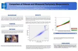
Correlation with tissue composition • High repeatability in measurement system • Good repeatability and correlation with elastic properties in phantom (normally a gel) or water Speed of sound Height of water column
But not so good in tissue .. Speed of sound Fat content (from chemical analysis)
Causes of error in samples Structure Shape and orientation Loading: 0.2% compressive strain - but hard to judge 0% strain Specimen preparation: Degassing – air bubbles have huge effect Region of muscle Region of fat (myosepta)
Velocities in other tissues • Important issue in ultrasound imaging • Fat composition very important • Data mixed; poor repeatability between different people/tissues • In-vivo the fat layer causes most distortion
Measuring shear velocity – the eye lens Oscilloscope Low frequency vibration excites shear wave Time of flight measurement gives velocity Pressure from motor? Time dependent effects?
For eye lens .. • High attenuation at ultrasound frequencies • Mechanical (or low frequency) wave excitation Results compare well with other estimates (spinning lens, deformation)
In-vivo methods • Can monitor the tendon/muscle etc in use and under different (real) loading • Limited in ultrasound windows • Signal may be affected by other tissue – eg fat layer • Possible to probe particular parts of the anatomy
Elastography • Ultrasound modality becoming standard • Designed for in-vivo use – used mainly in tumour detection • Measures tissue displacement – either through B-mode or r.f. image
Soft tissue biomechanicsElasticity imaging Sample Volume P = P0 Window Length P = P0+P … Beam Width v v Prof. Alison Noble
Measurements of tissue strain .. in-vivo • No absolute measure of length • Measure changes at different strains • Correlation of successive traces Displacement from strain (induced by temperature change in this case)
Strain estimation (from embedded heat source) Ultrasound image Strain estimation Based on coherent (r.f.) ultrasound data
Strain imaging – pilot study results Blue=high strain “ok” Red =low strain “suspect” Cancer Cyst Fibroadenoma DCIS Prof. Alison Noble
Tendon elastography Uses B-mode image; tracks speckle pattern Revell et al, IEEE Trans Medical Imaging, 24 6 2006 http://www.cs.bris.ac.uk/Research/Digitalmedia/cve/invivo.html
BUT .. • Inverse problem (local strain to elastic constants) very hard to solve • Effect of surrounding tissue • Orientation – limited number of ultrasound windows
Shear measurements • Generate a low frequency shear wave • Through differential movement • Through interference pattern from two transducers • From ‘pushing pulse’ • Watch propagation of wave with hgih frequency ultrasound
Shear measurements on musclesDifferential movement Muscle Shear modulus (relaxed) Shear modulus (contracted) Rectus femoris 5.87kPa 11.17kPa Biceps brachii 6.09 8.42 Hoyt et al, 2008
ARFI (Acoustic Radiation Force) imaging ‘Pushing pulse’ acting locally – can be high frequency for good focal volume control. Longitudinal wave Excites shear wave High speed image acquisition to capture shear velocity Tissue Adapted from Melodelima et al, Ultrasound in Medicine & BiologyVolume 32, Issue 3, March 2006, Pages 387-396 ‘pushing pulse’
Shearwave generation With thanks to Chris Haw, Alison Noble
Conclusions • Ex-vivo • Holding tissue – end effects? • Artificial loading conditions • Effect of neighbouring structure • In-vivo • Quantitative shear measurements • Displays of compression • Possibility of measuring under real loading • Limitation of viewing windows