Download
1 / 51
510 likes | 681 Views
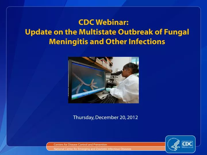
CDC Webinar: Update on the Multistate Outbreak of Fungal Meningitis and Other Infections. Centers for Disease Control and Prevention. National Center for Emerging and Zoonotic Infectious Diseases. Thursday, December 20, 2012. Agenda. Overview of new CDC Health Update

E N D
CDC Webinar:Update on the Multistate Outbreak of Fungal Meningitis and Other Infections Centers for Disease Control and Prevention National Center for Emerging and Zoonotic Infectious Diseases Thursday, December 20, 2012
Agenda • Overview of new CDC Health Update • Tom Chiller, MD, MPH (CDC) • Presentation of clinical case findings and strategies for treatment • AnuMalani, MD (St. Joseph’s Mercy Medical Center) • Michael Kasotakis, MD (St. Joseph’s Mercy Medical Center) • Q & A Session ***Please mute your phone during the presentations***
CDC Health Update • New data provides evidence that a substantial proportion of patients have developed localized infections following exposure to contaminated injections • Signs and symptoms of these spinal/paraspinal infections can be subtle and difficult to distinguish from the patient’s baseline chronic pain, clinicians should consider obtaining an MRI for patients with persistent but baseline symptoms • This decision should be made on a case-by-case basis after a discussion between the clinician and patient and taking into account the patient’s history regarding past and current symptoms More information online: http://www.cdc.gov/hai/outbreaks/meningitis.html
Fungal Infections Associated with Contaminated Methylprednisolone Injections — Spinal/Paraspinal Case Series Anurag Malani, M.D. Medical Director, Antimicrobial Stewardship Program Associate Medical Director, Infection Prevention & Control St. Joseph Mercy Health System – Ann Arbor Michael Kasotakis, M.D. Neuroradiology St. Joseph Mercy Health System – Ann Arbor December 20, 2012 4
Contents • Case 1: Parameningeal infection • Case 2: Epidural abscess • Case 3: Epidural abscess with minimal symptoms • Case 4: Osteomyelitis and discitis • Case 5: Sacroiliac osteomyelitis 5
Case 1: Parameningeal Infection Diagnosis: • Potential complication of fungal meningitis, despite treatment • MRI of epidural injection site indicated in meningitis • MRI results hard to interpret after surgery; may lag behind clinical improvement Treatment: • Prolonged amphotericin course in some arachnoiditis cases 7
Case 1: ParameningealInfectionHospitalization 1 • ID: 72 y/o female • Injection type: L3-L4 epidural • Chief complaint: 1 week of headache, neck stiffness, chills, and fatigue 15 days after injection • Lumbar puncture: confirmed meningitis • Medical treatment: • Inpatient: voriconazole + amphotericin for 5 days • Discharge: 26 days after injection • Symptoms: Improved • Outpatient treatment: oral voriconazole 8
Case 1: Parameningeal Infection Hospitalization 2 • Chief complaint: back pain, lower extremity weakness 36 days after injection • Magnetic Resonance Imaging (MRI): • Enhancement projecting intradural at L3 level 9
Case 1: MRI 36 Days after Injection Enhancement in dorsal thecal sac 10 Photo credit: St. Joseph Mercy Health Systems
Case 1: MRI 36 Days after Injection Enhancement and clumping of nerve roots. 11 Photo credit: St. Joseph Mercy Health Systems
Case 1: Parameningeal Infection Hospitalization 2 • Chief complaint: back pain, lower extremity weakness 36 days after injection • Magnetic Resonance Imaging (MRI): • Enhancement projecting intradural at L3 level • Operative treatment: • Procedure: evacuation of epidural abscess, L3 laminectomy • Intraoperative Findings: • Phlegmon-like material adherent to clumping nerve roots • Small amount of pus 12
Case 1: Operative Findings Thecal Sac with phlegmon 13 Photo credit: St. Joseph Mercy Health Systems
Case 1: Operative Pathology • Source: Intradural abscess • Stain: Hematoxylin and eosin (H+E) • Findings: purulent inflammation • Source: Intradural abscess • Stain: Gomori methenamine silver (GMS) • Findings: fungal hyphae 14 Photo credit: St. Joseph Mercy Health Systems
Case 1: Parameningeal Infection Hospitalization 2 • Medical treatment: • Inpatient: voriconazole + amphotericin for 5 weeks • MRI: 62 days after injection • Stable arachnoiditis with intradural involvement • Postoperative changes/enhancement at laminectomy site • Discharge: 71 days after injection • Symptoms: improved • Outpatient Treatment: oral voriconazole 15
Case 1: Parameningeal Infection —Summary Diagnosis: • Potential complication of fungal meningitis, despite treatment • MRI of epidural injection site indicated in meningitis • MRI results hard to interpret after surgery; may lag behind clinical improvement Treatment: • Prolonged amphotericin course in some arachnoiditis cases 16
Case 2: Epidural Abscess Diagnosis: • Potential rapid onset weeks after contaminated injection • Low MRI threshold in patients with persistent,worse, or new symptoms Treatment: • Neurosurgical consultation critical • Optimal treatment duration unknown 18
Case 2: Epidural Abscess • ID: 47 y/o male • Injection type: C6-C7 epidural • Chief complaint: headache, confusion, malaise, neck pain and spasms increasing since injection • Lumbar puncture: • 3 unremarkable studies, last 30 days after injection • MRI: • 3 unremarkable cervical studies, last 31 days after injection • 4th MRI 42 days after injection showed epidural abscess/phlegmon 19
Case 2: MRI- T1 Weighted 42 Days after Injection 31 Days after Injection C5-T1 phlegmon/epidural abscess 20 Photo credit: St. Joseph Mercy Health Systems
Case 2: MRI- T2 Weighted, Fat Suppressed 42 Days after Injection 31 Days after Injection C5-T1 phlegmon/epidural abscess 21
Case 2: Epidural Abscess • Operative treatment: • Procedure: evacuation of epidural abscess, C5-C7 laminectomy • Intraoperative findings: abscess and pus 22
Case 2: Operative Pathology • Source: Epidural abscess • Stain: H+E • Findings: acute inflammation next to bony spicule • Source: Epidural abscess • Stain: GMS • Findings: Rare fungal hyphae 23 Photo credit: St. Joseph Mercy Health Systems
Case 2: Epidural Abscess • Operative treatment: • Procedure: evacuation of epidural abscess, C5-C7 laminectomy • Intraoperative Findings: abscess and pus • Medical treatment: • Inpatient: voriconazole + amphotericin for 10 days • Discharge: 57 days after injection • Symptoms: improved • Outpatient treatment: oral voriconazole 24
Case 2: Epidural Abscess —Summary Diagnosis: • Potential rapid onset weeks after contaminated injection • Low MRI threshold in patients with persistent,worse, or new symptoms Treatment: • Neurosurgical consultation critical • Optimal treatment duration unknown 25
Case 3: Epidural Abscess with Minimal Symptoms Diagnosis: • Patients with minimal or baseline symptoms may harbor epidural infections • Low MRI threshold • Operative findings may not correlate with microbiology or pathology results 27
Case 3: Epidural Abscess with Minimal Symptoms • ID: 43 y/o female • Injection type: L3-L4, L4-L5 epidural 34 days apart • Chief complaint: Lower back pain similar to baseline (rated 3/10) • Lumbar puncture: 12 days after last injection • WBC 1 cell/µl • MRI: 41 days after last injection • Neuroforaminal enhancement L4-L5 • Enhancement at disc space margin 28
Case 3: MRI 41 Days after Last Injection Neuroforaminal enhancement L4-L5 Foraminal fat effaced Enhancement at disc space margin 29 Photo credit: St. Joseph Mercy Health Systems
Case 3: Epidural Abscess with Minimal Symptoms • Operative treatment: 44 days after last injection • Procedure: right L4-L5 laminectomy, medial facetectomy with foraminotomy of L4 root • Intraoperative findings: no abscess or pus • Anatomic Pathology: L4-L5 epidural fungal abscess 30
Case 3: Operative Pathology • Source: Paraspinal abscess • Stain: H+E • Findings: Acute inflammation with pigmented septate hyphae fragments 31 Photo credit: St. Joseph Mercy Health Systems
Case 3: Epidural Abscess with Minimal Symptoms • Operative treatment: 44 days after last injection • Procedure: right L4-L5 laminectomy, medial facetectomy with foraminotomy of L4 root • Intraoperative findings: no abscess or pus • Anatomic Pathology: L4-L5 epidural fungal abscess • Medical treatment: • Inpatient: voriconazole + amphotericin • Discharge: 52 days after last injection • Symptoms: improved • Outpatient treatment: oral voriconazole 32
Case 3: Epidural Abscess with Minimal Symptoms — Summary Diagnosis: • Patients with minimal or baseline symptoms may harbor epidural infections • Low MRI threshold • Operative findings may not correlate with microbiology or pathology results 33
Case 4: Osteomyelitis and Discitis Diagnosis: • Potential late complication of contaminated injection • Proactive outreach may identify patients earlier in their course Treatment: • Surgical consultation critical for suspected osteomyelitis 35
Case 4: Osteomyelitis and Discitis • ID: 28 y/o female • Injection type: L4-L5 epidural • Chief complaint: 3 weeks of left lower extremity weakness 90 days after injection • Lumbar puncture: approximately 70 days after injection • Normal • MRI: 91 days after injection • L4-L5 osteomyelitis and discitis • Paravertebral enhancement • L5 neuroforaminal disease 36
Case 4: MRI 91 Days after Injection L4-L5 osteomyelitis and discitis L5 neuroforaminal disease Enhancing disc margin 37 Photo credit: St. Joseph Mercy Health Systems
Case 4: MRI 91 Days after Injection Paravertebral enhancement 38 Photo credit: St. Joseph Mercy Health Systems
Case 4: Osteomyelitis and Discitis • Operative treatment: • Procedure: L4-L5microdiskectomy, irrigation and debridement of scar tissue on L5 nerve root • Pathology: fungal hyphae on GMS stain, chronic inflammation • Microbiology: mold, not yet speciated • Medical treatment: • Inpatient: voriconazole and amphotericin • Planned Discharge: 107 days after last injection • Symptoms: improving • Outpatient treatment: oral voriconazole 39
Case 4: Osteomyelitis and Discitis —Summary Diagnosis: • Potential late complication of contaminated injection • Proactive outreach may identify patients earlier in their course Treatment: • Surgical consultation critical for suspected osteomyelitis 40
Case 5: Sacroiliac Osteomyelitis Diagnosis: • Potential complications from contaminated sacroiliac injections • Low MRI threshold in patients with contaminated sacroiliac injections Treatment: • Unclear criteria for surgical intervention 42
Case 5: Sacroiliac Osteomyelitis • ID: 60 y/o female • Injection type: right sacroiliac joint • Chief complaint: progressive right sided pain, difficulty ambulating • MRI: 44 days after injection • Right sacroiliac joint septic arthritis • Adjacent osteomyelitis in inferior aspect of joint 43
Case 5: MRI 44 Days after Injection Right sacroiliac joint septic arthritis with adjacent osteomyelitis in inferior aspect of the joint Fat suppressed post contrast 44 Photo credit: St. Joseph Mercy Health Systems
Case 5: Sacroiliac Osteomyelitis • Operative treatment: • Procedure: incision and drainage • Intraoperative findings: small amount of purulent fluid 45
Case 5: Operative Pathology • Source: sacroiliac biopsy • Stain: H+E • Findings: neutrophil and plasma cell infiltration next to bony spicules, consistent with acute osteomyelitis 46 Photo credit: St. Joseph Mercy Health Systems
Case 5: Sacroiliac Osteomyelitis • Operative treatment: • Procedure: incision and drainage • Intraoperative findings: small amount of purulent fluid • Pathology: acute osteomyelitis of right posterior ilium • Microbiology: Exserohilum confirmed • Medical treatment: • Inpatient: intravenous voriconazole • Discharge: 58 days after last injection • Symptoms: improved • Outpatient treatment: oral voriconazole 47
Case 5: Sacroiliac Osteomyelitis —Summary Diagnosis: • Potential complications from contaminated sacroiliac injections • Low MRI threshold in patients with contaminated sacroiliac injections Treatment: • Unclear criteria for surgical intervention 48
Conclusions • Parameningeal infections, epidural abscess, osteomyelitis with discitis, and sacroiliac osteomyelitis are potential complications of contaminated methylprednisolone injections • Proactive outreach may identify patients earlier in their course • Maintain low threshold to order MRI with and without contrast at the spinal or paraspinal injection site in patients with persistent, worse, or new symptoms • Early surgical consultation essential for management of spinal and paraspinal infections • Optimal duration of medical treatment not yet known 49
Acknowledgements St. Joseph Mercy Health Systems Ann Arbor Paul Valenstein, MD Centers for Disease Control and Prevention Mycotic Diseases Branch Tom Chiller, MD MPH Monika Roy, MD Division of Healthcare Quality Promotion John Jernigan, MD Alice Guh, MD MPH Raymund Dantes, MD MPH 50