Download
1 / 1
10 likes | 128 Views
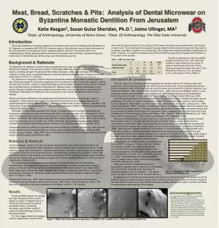
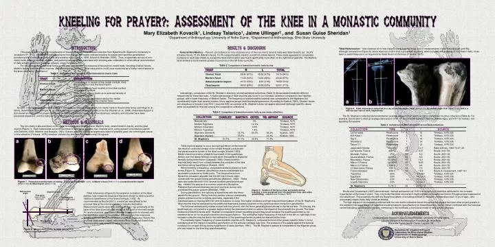
Mary Elizabeth Kovacik 1 , Lindsay Talarico 1 , Jaime Ullinger 2 , and Susan Guise Sheridan 1 1 Department of Anthropology, University of Notre Dame; 2 Department of Anthropology, Ohio State University.

E N D
Mary Elizabeth Kovacik1, Lindsay Talarico1, Jaime Ullinger2 , and Susan Guise Sheridan1 1Department of Anthropology, University of Notre Dame; 2Department of Anthropology, Ohio State University Tibial Retroversion:Inter-observer error was insignificant across the three sets of measurements of this feature (n=25; p≤0.59). Although retroversion (Figure 4) alone does not confirm that a group was squatting, when included with a variety of non-metric traits, it has been a useful measure in an argument for deep flexion of the knee (Trinkaus, 1975). This study is part of a larger consideration of kneeling behavior in a skeletal collection from Byzantine St. Stephen’s monastery in Jerusalem (5th-7th C). Historical and liturgical texts from the site and region indicate kneeling for prayer and repetitive genuflection (numbering the hundreds) as part of the daily worship cycle (Bautch, 1999; Driscoll & Sheridan, 2000). Thus, a systematic survey of non-metric traits, musculo-skeletal markers, and pathological responses associated with kneeling was undertaken in a biocultural reconstruction of daily activity patterns in this large, affluent, urban monastery. For the current study, the distal femur (n=99) was analyzed for the presence of several non-metric traits, including Charles’ facets, Martin’s facets, osteochondritic imprints, and tibial imprints (Table 1). Tibial retroversion (n=25) was measured as a further test of stress on the knee induced by hyperflexion. Femoral Non-Metrics -- Percent concordance for inter-observer error of the non-metric femoral traits was determined to be: 93.9% (Charles’ facet), 77.4% (Martin’s facet), 79.1% (osteochondritic imprint), and 87.5% (tibial imprint). These traits appeared in comparable numbers on each side (Table 2), except the tibial imprint which was found significantly more often on the right femur (p≤0.05). The Martin’s facet showed a trend towards greater occurrence on the left femur (p=0.06). Table 1. Definitions and locations of the femoral non-metric traits TRAIT DESCRIPTION COLLECTION CHARLES’ OSTEO. TIB. IMPRINT SOURCE Charles’ Facet Located near or as part of the medial condyle; divot-like impression near the tibial imprint Neandertals - 0% - Trinkaus, 1975 Interestingly, comparison of the St. Stephen’s results to several published collections (Table 3) demonstrated markedly different frequencies for these two traits. A higher percentage of tibial imprints was found in our monastic collection compared to two Nigerian groups, with a lower frequency of Martin’s facets. The frequency of osteochondritic imprints was comparable to the Nigerians, but considerably higher than several modern Homo sapiens groups and Neandertal specimens. According to Kostick (1963), Charles’ facets are ubiquitous in femurs (“over 90%”), thus the 93% occurrence at St. Stephen’s does not appear abnormal (although specific values were not available for this trait among the comparative collections). Modern Egyptians - - 12.1% - Trinkaus, 1975 Martin’s Facet Crescent-shaped facet located on trochlear surface of the lateral condyle Modern Icelanders - 4.6% - Trinkaus, 1975 Osteochondritic Imprint Hole, cavity, or bony build-up on articular lamella of the lateral condyle Modern Yugoslavs - 1.4% - Trinkaus, 1975 Nigerians (Series A) - 78.7% 25.3% 39.2% Kostick, 1963 Table 3. Comparison of femoral non-metric traits across study collections Tibial Imprint Thumb print-like impression above the medial condyle; may border the Charles’ facet Nigerians (Ibadan) - 75.9% - 33.3% Kostick, 1963 As seen in the sequence of related posters in this session, when combined with non-metric traits of the proximal femur (Jennings et. al., 2004), distal tibia & talus (Ullinger, et. al, 2004), as well as musculoskeletal markers of the hip (Hayden et. al., 2004), these data support a biocultural reconstruction of prayer practice at St. Stephen’s (related analyses of the calcaneus, vertebra, and shoulder have been presented elsewhere, and the hallux is currently under investigation). St. Stephen’s 93.7% 57.1% 30.5% 71.4% MARTIN’S Figure 4.Tibial retroversion measured on x-ray: a) extreme angle = 52.0°[EBND 5.02];b) minimal angle 15.9° [EBND 8.162];and c) a macroscopic view of each, respectively. - The St. Stephen’s collection demonstrated an average angle of 27.5° which is high in comparison to other collections (Table 4). For example, Boulle (2001) cited an average retroversion of 14-17° for a French collection from the Middle Ages, and ≤11° for modern, non-squatting Europeans. b a c - - a b c d The non-metric traits examined on the distal femur included Charles’ facet, Martin's facet, osteochondritic imprint, and the tibial imprint (Figure 1). Each feature was scored three times to determine possible inter-observer error, using percent concordance (Jacobi and Danforth, 2002; Waldron and Rodgers, 1991). A direct analysis of bilateral asymmetry was not possible given the commingled nature of the collection (Trinkaus, 1978); however, variation by side was calculated using Chi-square. Table 4. Comparison of tibial retroversion across study collections COLLECTION TIME n ° SOURCE 1 La Ferrassie Pleistocene 15 Trinkaus, 1975:335 1 Kiik-Koba 1 Pleistocene 16 Trinkaus, 1975:335 Tibial imprints appear to occur during hyperflexion of the knee as the result of continued contact of the medial femoral condyle with the lateral posterior border of the tibial condyle (Kostick 1963). Martin’s facet have been related to movement of the quadriceps tendon over the lateral femoral condyle when the patella is displaced laterally during knee flexion (Capasso 1999). Osteochondritic imprints likely result from contact between the condyles of the tibia and femur during hyperflexion (Kostick, 1963) When kneeling or squatting, comparable stress is placed on both knees (Figure 3). However, genuflection exerts considerable but asymmetric pressure on these joints. The liturgical texts from Byzantine Jerusalem stipulate that the left knee should make contact with the ground during genuflection (Baldovin, 1992). These texts also prescribe from 50 to several hundred genuflections per day. Indeed, this was such a common practice in Byzantine Palestine that several observances (such as those during Lent) prohibited this prayer posture (Sheridan, 1999). During genuflection, the right leg is hyperflexed with the femur parallel to the ground. Rising from this position places considerably greater pressure on the condyles (and toes), especially if the 1 Spy 2 Pleistocene 18 Trinkaus, 1975:335 1 Shanidar 2 Pleistocene 14 Trinkaus, 1975:335 1 Tabun C1 Pleistocene 12 Trinkaus, 1975:335 46 La Favorite, France 1-2nd C 9.57 Boulle, 2001:53 Figure 1. Femoral non-metric traits,including: a) Charles’ facet[EBND 1.272];b) Martin’s facet[EBND 3.117];c) osteochondritic imprint[EBND 5.160];d) tibial imprint[EBND 6.132]. Figure 3. Relation of the femur, tibia, and patella during kneeling, a more sustained form of hyperflexion than seen when genuflecting.Adapted from Trinkaus (1975). Boulle and Coussement (1997) demonstrated marked retroversion at 19.8º in a collection of fetal tibia, attributed to the constant hyperflexion of the knee in utero. They concluded that tibial retroversion might indicate continued knee flexion throughout post-natal period into adulthood. This possibility fits with the Byzantine pattern of including children in monastic communities (over 10 yrs of age) , who presumably began these daily rituals as oblates. The high degree of retroversion, combined with the non-metric indicators found throughout the leg and foot (see other project posters in this session) indicate a pattern of repetitive kneeling behavior (genuflection) for these Byzantine monks. When combined with the historical record indicating genuflections regularly numbering in the hundreds, a biocultural model of daily activity emerges. Tibial retroversion (Figure 2), the posterior inclination of the tibial plateau, was measured because it has been shown to have a positive relation to hyperflexion of the knee (Trinkaus, 1975). As recommended by Boulle (2001), a metal bar was affixed to the proximal tibia at the internal plateau to indicate declination. Measurement points were then marked on the x-ray, proximally at the base of the tibial tuberosity and distally at the point of minimal breadth. An ‘anatomical axis’ was drawn between these points to intersect with the declination angle at the plateau. A straight line then intersects these points from which retroversion angle was measured. Twenty-five proximal tibiae were x-rayed; retroversion was measured three times. Reproducibility was compared using Students’ t-test. 55 Michelet, France 4th C 8.2 Boulle, 2001:53 Retroversion angle Anatomical axis Table 2. Comparison of femoral non-metric traits by side 42 Launa-Mollard, France 6-8th C 8.6 Boulle, 2001:53 individual leans on that leg with their arm for balance in rising. The higher incidence and right side-dominant pattern of the St. Stephen’s tibial imprints may be explained by the additional downward pressure exerted on the right knee when rising from genuflection. First point (base of tibial tuberosity) 42 Mountalieu, France 10-12th C 10.1 Boulle, 2001:53 R L TOTAL TRAIT Second point (minimum breadth) 20 Mâcon I, France 14-16th C 8.05 Boulle, 2001:53 The left knee subsequently makes contact with the ground, with the femur generally perpendicular to the flat surface. For this leg, the quadriceps are contracted to a greater degree during the downward motion to reduce the impact of the knee with the ground. The lower incidence of Martin’s facets at St. Stephen’s compared to known squatting groups may result because the quadriceps tendon is not stretched as far (or for as great a duration) during genuflection. The somewhat higher frequency of this trait in the left vs. right legs of our monastic collection may be due to the contraction of the quadriceps tendon to slow the descent of the knee. The markedly higher frequency of osteochondritic imprints at St. Stephen’s, compared to most of the groups listed in Table 3, is not surprising given the impact of repetitive genuflection on the articular cartilage over time. The pressure of the patella against the femoral condyles can exceed 400 kg during hyperflexion (Calais-Germain, 1991). The St. Stephen’s pattern is comparable to the Nigerian group, who are noted for the time they spend squatting. Charles’ Facet 39/43 (91%) 35/36 (97%) 74/79 (94%) 17 Mâcon II, France 18th C 8.4 Boulle, 2001:53 Martin’s Facet 11/22 (50%) 13/20 (65%) 24/42 (57%) 44 L’Observance, France 18th C 7.3 Boulle, 2001:53 56 France (fetuses) 20th C 19.8 Boulle & Coussement, 1997:191 Osteochondritic Imprint 10/33 (30%) 8/26 (31%) 18/59 (31%) - Yugoslavs 20th 9.4±3.7 Trinkaus, 1975:335 Tibial Imprint 45/53 (85%) 20/38 (53%) 65/91 (71%) - Egyptians 20th 14.7±3.2 Trinkaus, 1975:335 Undergraduate Research Opportunities Program, University of Notre Dame L’École Biblique et Archéologique Française de Jérusalem Blairsville Family Health Center, Latrobe, PA - New Caledonians 20th 13.4±3.9 Trinkaus, 1975:335 15 Japanese/Chinese Neolithic 14.7 Baba & Endo, 1982:T4.27, 28 - Loyalty Islanders 20th 14.9±3.1 Trinkaus, 1975:335 Tibial plateau (metal axis) 24 St. Stephen’s 5-7th C 27.5±7.3 Figure 2.Measurement points for tibial retroversion. Adapted from Boulle (2001) Byzantine St. Stephen’s Project Laboratory for Biocultural Studies Department of Anthropology University of Notre Dame .