Download
1 / 73
740 likes | 990 Views
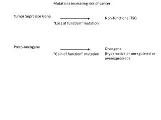
APC Beta Catenin pathway Belongs to tumor suppressor gene Functions It is a cytoplasmic protein whose dominant function is to regulate the intracellular levels of β- catenin , a protein with many functions. Down regulation of growth promoting signals.

E N D
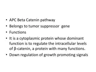
APC Beta Catenin pathway • Belongs to tumor suppressor gene • Functions • It is a cytoplasmic protein whose dominant function is to regulate the intracellular levels of β-catenin, a protein with many functions. • Down regulation of growth promoting signals
Germ line mutations at 5q21 loci associated with Familial APC in which all individuals born with single mutated allele develop polys and 1 or 2 malignant transform in their 20s leads to colon ca • These patients consistently show loss of a tumor suppressor gene APC
β-catenin binds to the cytoplasmic portion of E-cadherin, a cell surface protein that mediates intercellular interactions; • it can translocate to the nucleus and activate cell proliferation. β-catenin is an important component of the so-called WNT signaling pathway that regulates cell proliferation WNT is a soluble factor that can induce cellular proliferation
In quiescent cells, which are not exposed to WNT, cytoplasmic β-catenin is degraded by a destruction complex, of which APC is an integral part • With loss of APC (in malignant cells), β-catenin degradation is prevented, and the WNT signaling response is inappropriately activated in the absence of WNT • This leads to transcription of growth-promoting genes, such as cyclin D1 and MYC.
NF1 and NF2 • The person who inherit single mutated allele develop numerous benign neurofibromas and optic nerve gliomas and leads to inactivation of 2nd copy called neurofibromatosis type 1 • Later develop into malignant peripheral nerve sheet tumor
Germ line muatations in NF2 leads to Neurofibromatosis type 2
Evasion of Apoptosis • Accumulation of neoplastic cells may result • from activation of growth-promoting oncogenes • or inactivation of growth-suppressing tumor suppressor genes, • also from mutations in the genes that regulate apoptosis
The pro-apoptotic proteins, BAX and BAK, are required for apoptosis and directly promote mitochondrial permeabilization • anti-apoptotic members of this family BCL2 and BCL-XL • proteins (so-called BH3-only proteins) including BAD, BID, and PUMA, regulate the balance between the pro- and anti-apoptotic members of the BCL2 family
BAX and BAK are activated and form pores in the mitochondrial membrane. Cytochromec leaks into the cytosol, where it binds to APAF-1, activating caspase 9. Like caspase 8 of the extrinsic pathway, caspase 9 can cleave and activate the executioner caspases.
The causes for evasion of apoptosis in tumor cells due to • 1.reduced levels of CD95 may render the tumor cells less susceptible to apoptosis by Fasligand (FasL). • 2.Some tumors have high levels of FLIP, a protein that can bind death-inducing signaling complex and prevent activation of caspase 8.
3. best established is the role of BCL2 in protecting tumor cells from apoptosis • Why lymphomas are slow growing? • There is overexpression of BCL2 proteins • Inturn increases BCL2/BCL-XL buffer, protecting lymphocytes from apoptosis and allowing them to survive for long periods; there is therefore a steady accumulation of B lymphocytes
resulting in lymphadenopathy and marrow infiltration. Because BCL2-overexpressing lymphomas arise in large part from reduced cell death rather than explosive cell proliferation, they tend to be indolent (slow growing) compared with many other lymphoma • 85% of follicular B-cell lymphomas the anti-apoptotic gene BCL2 is activated by the t(8;14) translocation.
Limitless Replicative Potential • normal human cells have a capacity of 60 to 70 doublings. • cells lose the capacity to divide and enter senescence • shortening of telomeres at the ends of chromosomes. short telomeres seem to be recognized by the DNA repair machinery as double-stranded DNA breaks, and this leads to cell cycle arrest mediated by p53 and RB. Cells in which the checkpoints are disabled by p53 or RB mutations
short telomeres seem to be recognized by the DNA repair machinery as double-stranded DNA breaks, and this leads to cell cycle arrest mediated by p53 and RB.Cells in which the checkpoints are disabled by p53 or RB mutations
the nonhomologous end-joining pathway is activated to save the cell, joining the shortened ends of two chromosomes. This inappropriately activated repair system results in dicentric chromosomes that are pulled apart at anaphase, resulting in new double-stranded DNA breaks. The resulting genomic instability from the repeated bridge-fusion-breakage cycles eventually produces mitotic catastrophe, characterized by massive cell death.
Tumor cells must also develop ways to avoid both cellular senescence and mitotic catastrophe If during crisis a cell manages to reactivate telomerase, the bridge-fusion-breakage cycles cease and the cell is able to avoid death. • However, during this period of genomic instability that precedes telomerase activation, numerous mutations could accumulate, helping the cell march toward malignancy. Passage through a period of genomic instability probably explains the complex karyotypes frequently seen in human carcinomas
telomere maintenance is seen in virtually all types of cancers. • In 85% to 95% of cancers, this is due to up-regulation of the enzyme telomerase. A • A few tumors use other mechanisms, termed alternative lengthening of telomeres, which probably depend on DNA recombination. • The progression from colonic adenoma to colonic adenocarcinoma, early lesions had a high degree of genomic instability with low telomerase expression.
Development of Sustained Angiogenesis • tumors cannot enlarge beyond 1 to 2 mm in diameter unless they are vascularized • tumors require delivery of oxygen and nutrients and removal of waste products
neo-angiogenesis In a tumor • Formation of new vessel from previously existing vessel • Early in their growth, most human tumors do not induce angiogenesis. • They remain small or in situ for years
Angiogenesis involves increased production of angiogenic factors and/or loss of angiogenesis inhibitors. • These factors may be produced directly by the tumor cells themselves or by inflammatory cells (e.g., macrophages) or other stromal cells associated with the tumors.
Stimuli, is hypoxia • Factors in angiogenesis are • 1.vascular endothelial growth factor (VEGF) • 2.Hypoxia-induced factor-1α (HIF1α) • 3.von Hippel-Lindau protein
(VHL syndrome) • VHL gene are associated with • 1. Hereditary renal cell cancers, 2.pheochromocytomas, • 3.Hemangiomas of the central nervous system, • 4. Retinal angiomas, and • 5. Renal cysts
What is VHL syndrome? • VHL acts as a tumor suppressor gene,in the process of angiogenesis and germ-line mutations of the VHL gene are called as VHL syndrome.
In normal cells, p53 can stimulate expression of anti-angiogenic molecules, thrombospondin-1, and repress expression of pro-angiogenic molecules, VEGF. • Loss of p53 in tumor cells favor the envirnoment for angiogenesis. • Mutations of RAS or MYC up-regulate the production of VEGF
Proteases, either elaborated by the tumor cells directly or from stromal cells in response to the tumor, are also involved in regulating the balance between angiogenic and anti-angiogenic factors. • proteases can release the angiogenic basic FGF stored in the extracellular matrix (ECM);
three potent angiogenesis inhibitors-angiostatin, • endostatin, and • vasculostatin-are produced by proteolytic cleavage of plasminogen, collagen, and transthyretin, respectively. • anti-angiogenesis therapy. anti-VEGF antibody is now approved for the treatment of several types of cancers
Ability to Invade and Metastasize • subdivided into two phases: invasion of ECM and • vascular dissemination, and homing of tumor cells
Invasion of Extracellular Matrix (ECM) • Invasion of the ECM is an active process that requires four steps • 1.Detachment of tumor cells from each other • 2.Degradation of ECM • 3.Attachment to novel ECM components • 4.Migration of tumor cells
TRANSFORMATIONGROWTHBM INVASIONANGIOGENESISINTRAVASATIONEMBOLIZATIONADHESIONEXTRAVASATIONMETASTATIC GROWTHetc.
Human tissues are organized into a series of compartments separated from each other by two types of ECM: basement membranes and interstitial connective tissue • ECM is composed of collagens, glycoproteins, and proteoglycans.
A carcinoma first must breach the underlying basement membrane, • then traverse the interstitial connective tissue, and • ultimately gain access to the circulation by penetrating the vascular basement membrane
Invasion of the ECM is an active process that requires four steps • Detachment of tumor cells from each other • Degradation of ECM • Attachment to novel ECM components • Migration of tumor cells
The first step in the metastatic cascade is a loosening of tumor cellsE-cadherins act as intercellular glues, and their cytoplasmic portions bind to β-catenin). Adjacent E-cadherin molecules keep the cells together; in addition, E-cadherin can transmit antigrowth signals by sequestering β-catenin. • E--cadherin function is lost in almost all epithelial cancers, either by mutational inactivation of E-cadherin genes
inappropriate expression of the SNAIL and TWIST transcription factors, which suppress E-cadherin expression. • 2.invasion is local degradation of the basement membrane and interstitial connective tissue. Tumor cells may either secrete proteolytic enzymes themselves or induce stromal cells to elaborate proteases.
matrix metalloproteinases (MMPs), cathepsin D, and urokinaseplasminogen activator, have been implicated in tumor cell invasion. • MMPs regulate tumor invasion by releasing ECM-sequestered growth factors. Indeed, cleavage products of collagen and proteoglycans also have chemotactic, angiogenic, and growth-promoting effects
, MMP-9 is a gelatinase that cleaves type IV collagen of the epithelial and vascular basement membrane and also stimulates release of VEGF from ECM-sequestered pools • protease inhibitors as therapeutic agents.? • MMP inhibitors are decreased lads to favor the tumor invasion.
3.Attachment of tumor cells to ECM proteins • 4.Locomotion is the final step of invasion, propelling tumor cells through the degraded basement membranes and zones of matrix proteolysis.
tumor cell-derived cytokines, some growth factors (e.g., insulin-like growth factors I and II) have chemotactic activity for tumor cells. • Stromal cells also produce paracrine effectors of cell motility, such as hepatocyte growth factor/scatter factor (HGF/SCF), which bind to receptors on tumor cells. Concentrations of HGF/SCF are elevated at the advancing edges of the highly invasive brain tumor glioblastomamultiforme, supporting their role in motility.
. Concentrations of HGF/SCF are elevated at the advancing edges of the highly invasive brain tumor glioblastomamultiforme, supporting their role in motility
Vascular Dissemination and Homing of Tumor Cells • When in the circulation, tumor cells are vulnerable to destruction by host immune cells • In the bloodstream, some tumor cells form emboli by aggregating and adhering to circulating leukocytes, particularly platelets; aggregated tumor cells are thus afforded some protection from the antitumor host effector cells.
Genomic Instability-Enabler of Malignancy • the ability of normal cells to repair DNA damage. The importance of DNA repair in maintaining the integrity of the genome • Inherited disorders in which genes that encode proteins involved in DNA repair are defective. Individuals born with such inherited defects in DNA repair proteins are at a greatly increased risk of developing cancer.
Hereditary Nonpolyposis Colon Cancer Syndrome • The defect in DNA repair genes • This disorder, characterized by familial carcinomas of the colon affecting predominantly the cecum and proximal colon results from defects in genes involved in DNA mismatch repair. • DNA repair genes behave like tumor suppressor genes
XerodermaPigmentosum Patients, are at increased risk for the development of cancers of the skin exposed to the ultraviolet (UV) light contained in sun rays. The basis of this disorder is defective DNA repair. UV light causes cross-linking of pyrimidine residues, preventing normal DNA replication.
MicroRNAs (MiRNAs) and Cancer • miRNAs control cell growth, differentiation, and cell survival,
MicroRNAs (MiRNAs) and Cancer • miRNAs control cell growth, differentiation, and cell survival, • miRNAs can participate in neoplastic transformation either by increasing the expression of oncogenes or • reducing the expression of tumor suppressor genes.
Uses of Micro RNA • 1.Drugs that inhibit or augment the functions of miRNAs could be useful in chemotherapy. Since miRNAs regulate normal cellular • 2.Differentiation, the patterns of miRNA expression ("miRNA profiling") can provide clues to the cell of origin and classification of tumors.
ETIOLOGY OF CANCER: CARCINOGENIC AGENTS • 1) Chemicals, • (2) Radiant energy, and • (3) Microbial agents.
Chemical Carcinogens • Direct-Acting Agents • Indirect-Acting Agents
Direct-Acting Carcinogens • ALKYLATING AGENTS • Anticancer drugs (cyclophosphamide, chlorambucil, nitrosoureas, and others • β-Propiolactone • POLYCYCLIC AND HETEROCYCLIC AROMATIC HYDROCARBONS
AROMATIC AMINES, AMIDES, AZO DYES • Natural Plant and Microbial Products • Aflatoxin B1 • Griseofulvin • Betel nuts • OTHERS • Nitrosamine and amides Vinyl chloride, nickel, chromium Insecticides, fungicides