Download
1 / 1
20 likes | 211 Views
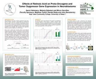
Effects of Retinoic Acid on Proto-Oncogene and Tumor Suppressor Gene Expression in Neuroblastoma. Aaron Halemano, Melanie Nakatani and Micon Garvilles (Faculty Sponsors: Matthew Tuthill, Randal Wada and John Berestecky) Kapi ‘ olani Community College, University of Hawai ‘ i. INTRODUCTION

E N D
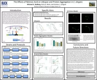
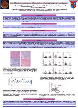
Effects of Retinoic Acid on Proto-Oncogene and Tumor Suppressor Gene Expression in Neuroblastoma Aaron Halemano, Melanie Nakatani and Micon Garvilles (Faculty Sponsors: Matthew Tuthill, Randal Wada and John Berestecky) Kapi‘olani Community College, University of Hawai‘i INTRODUCTION Neuroblastoma is the most common extracranial solid tumor found in children, resulting in approximately six hundred and fifty new cases in the U.S. annually. This peripheral nervous system cancer is derived from pluripotent stem cells that arise out of the neural crest during development. N-myc oncogene overexpression and/or amplification is often predictive of poor prognosis, and correlates with advanced and aggressive disease, as well as resistance to chemotherapy. More recent clinical trials involving retinoic acid adjuvant therapy have shown efficacy in the setting of minimal residual disease. Numerous laboratory studies have also shown that retinoic acid induces the downregulation of N-myc proto-oncogene expression in select neuroblastoma cell lines. While the effects of retinoic acid appear to be mediated through promoter regulation of the N-myc gene, the exact mechanism of N-myc downregulation has not been clearly established. The specific aim of this study is to determine if possibly the expression of other gene targets are also modulated by retinoic acid. RESULTS As expected, retinoic acid treatment of the LA-N-5 neuroblastoma cell line resulted in little change in HPRT1 reference gene expression. Moreover, transcriptional activity of the positive control N-myc gene was downregulated. However, retinoic acid treatment of LA-N-5 cells resulted in either modest, or significant upregulation of the target genes RB1, AUTS2 and ROR1. CONCLUSIONS Our studies continue in order to determine if the observed changes are both reproducible in other neuroblastoma cell lines and significant enough to potentially have a physiological effect. Moreover, we are investigating if the effects of retinoic acid are enhanced with concurrent treatment of a cyclin-dependent kinase 4/6 inhibitor. The culmination of which may allow for a more an alternate treatment regimen at lower individual concentrations of each drug, as well as more conclusively determine if these genes are modulated by retinoic acid. FUTURE PLANS Polymerase chain reaction will be used to amplify target gene fragments from LA-N-5 (a). These amplicons will then be TA-cloned into the pDrive vector (b), transformed into E. coli (c) and screened for ampicillin resistant white colonies (d). The positive clones will then be confirmed via polymerase chain reaction and validated via DNA sequencing. Successful clones will be used to generate standard curves for subsequent absolute quantification real-time polymerase chain reactions. (a) (b) (c) (d) (e) (a) (b) (c) (d) Figure 2. Real- time polymerase chain reaction for HPRT1, N-myc and RB1. Amplification plot (a). ddCt normalized fold expression (b). Melt peak chart of HPRT1 (c), N-myc (d), and RB1 (e). (a) (b) METHODOLOGY The LA-N-5 human neuroblastoma cell line was treated for four days with retinoic acid or dimethyl sulfoxide (control medium) before total RNA extraction. Quantitative real-time polymerase chain reaction analyses were then performed against four gene targets: RB1, TP53, AUTS2 and ROR1. N-myc downregulation via retinoic acid served as the positive control, while the HPRT1 (hypoxanthine-guanine phosphoribosyltransferase) gene target served as the reference gene and allowed for the determination of relative levels of gene expression across different sample sets. ACKNOWLEDGEMENTS We thank A. Garcia, C. Allen, S. Kawasaki, K. Noji and K. Noa for their ongoing laboratory and administrative support. Funding for this project was provided by the Kapiolani Community College STEM Program, the Perkins Grant and the National Center for Research Resources (5P20RR016467-11) and the National Institute of General Medical Sciences (8 P20 GM 103466) from the National Institutes of Health. The University of Hawaii RMATRIX Program is supported by award number U545MD007584 from the National Institute on Minority Health and Health Disparities, National Institutes of Health. The content is solely the responsibility of the authors and does not represent the official views of the National Institutes of Health. (c) (d) (e) LITERATURE CITED 1. S. Toegel, W. Huang, C. Piana, F. M. Unger, M. Wirth, M. B. Goldring, F. Gabor and H. Viernstein. 2007. Selection of reliable reference genes for qPCR studies on chondroprotective action. BMC Molecular Biology, 8:13. 2. Y. Liu and W.F. Bodmer.2005. Analysis of P53 mutations and their expression in 56 colorectal cancer cell lines. PNAS. 103:4, 976-981. 3. M. Hudecek, T. M. Schmitt, S. Baskar, M.T. Lupo-Stanghellini, T. Nishida, T. N. Yamamoto, M. Bleakley, C. J. Turtle, W. C. Chang, H. A. Greisman, B. Wood, D. G. Maloney, M. C. Jensen, C. Rader and S. R. Riddell. 2010. The B-cell tumor-associated antigen ROR1 can be targeted with T cells modified to express a ROR1-sepcific chimeric antigen receptor. Blood, 116:22, 4532-4541. 4. RTPrimerDB ID:1194 = Nmyc. 5. RTPrimerDB ID:8038 = RB1. Figure 3. Real-time polymerase chain reaction for HPRT1, AUTS2 and ROR1. Amplification plot (a). ddCt normalized fold expression (b). Melt peak chart of HPRT1 (c), AUTS2 (d), and ROR1 (e). Figure 1. Workflow methodology. LA-N-5 neuroblastoma cells were treated with retinoic acid, total RNA was then extracted and served as template for RT-PCR analyses.