Download
1 / 30
300 likes | 469 Views
Muscles and Muscle Tissue. 9. Part A. Muscle Overview. The three types of muscle tissue are skeletal, cardiac, and smooth These types differ in structure, location, function, and means of activation. Muscle Similarities.

E N D
Muscles and Muscle Tissue 9 Part A
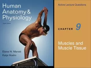
Muscle Overview • The three types of muscle tissue are skeletal, cardiac, and smooth • These types differ in structure, location, function, and means of activation
Muscle Similarities • Skeletal and smooth muscle cells are elongated and are called muscle fibers • Muscle contraction depends on two kinds of myofilaments – actin and myosin • Muscle terminology is similar • Sarcolemma – muscle plasma membrane • Sarcoplasm – cytoplasm of a muscle cell • Prefixes – myo, mys, and sarco all refer to muscle
Types of Muscle Tissue • Skeletal Muscle Tissue • Cardiac Muscle Tissue • Smooth Muscle Tissue
Functional Characteristics of Muscle Tissue • Excitability, or irritability – the ability to receive and respond to stimuli • Contractility – the ability to shorten forcibly • Extensibility – the ability to be stretched or extended • Elasticity – the ability to recoil and resume the original resting length
Muscle Function • Skeletal muscles are responsible for all locomotion • Cardiac muscle is responsible for coursing the blood through the body • Smooth muscle helps maintain blood pressure, and squeezes or propels substances (i.e., food, feces) through organs • Muscles also maintain posture, stabilize joints, and generate heat
Skeletal Muscle: Nerve and Blood Supply • Each muscle is served by one nerve, an artery, and one or more veins • Each skeletal muscle fiber is supplied with a nerve ending that controls contraction • Contracting fibers require continuous delivery of oxygen and nutrients via arteries • Wastes must be removed via veins
Myofibrils Figure 9.3 (b)
Sarcomeres • The smallest contractile unit of a muscle • The region of a myofibril between two successive Z discs • Composed of myofilaments made up of contractile proteins • Myofilaments are of two types – thick and thin
Sarcomeres Figure 9.3 (c)
Myofilaments: Banding Pattern • Thick filaments – extend the entire length of an A band • Thin filaments – extend across the I band and partway into the A band • Z-disc – coin-shaped sheet of proteins (connectins) that anchors the thin filaments and connects myofibrils to one another
Myofilaments: Banding Pattern (Contain desmin protein) Figure 9.3 (c, d)
Ultrastructure of Myofilaments: Thick Filaments • Thick filaments are composed of the protein myosin • Each myosin molecule has a rodlike tail and two globular heads • Tails – two interwoven, heavy polypeptide chains • Heads – two smaller, light polypeptide chains called cross bridges
Ultrastructure of Myofilaments: Thick Filaments Figure 9.4 (a)(b)
Ultrastructure of Myofilaments: Thin Filaments • Thin filaments are chiefly composed of the protein actin • Each actin molecule is a helical polymer of globular subunits called G actin • The subunits contain the active sites to which myosin heads attach during contraction • Tropomyosin and troponin are regulatory subunits bound to actin
Ultrastructure of Myofilaments: Thin Filaments Figure 9.4 (c)
Arrangement of the Filaments in a Sarcomere • Longitudinal section within one sarcomere Figure 9.4 (d)
Sarcoplasmic Reticulum (SR) • SR is an elaborate, smooth endoplasmic reticulum that mostly runs longitudinally and surrounds each myofibril • Functions in the regulation of intracellular calcium levels • T tubules associate with the paired terminal cisternae to form triads
Sarcoplasmic Reticulum (SR) Figure 9.5
T Tubules • T tubules are continuous with the sarcolemma • They conduct impulses to the deepest regions of the muscle • These impulses signal for the release of Ca2+ from adjacent terminal cisternae
Sliding Filament Model of Contraction • Thin filaments slide past the thick ones so that the actin and myosin filaments overlap to a greater degree • In the relaxed state, thin and thick filaments overlap only slightly • Upon stimulation, myosin heads bind to actin and sliding begins • Each myosin head binds and detaches several times during contraction, acting like a ratchet to generate tension and propel the thin filaments to the center of the sarcomere • As this event occurs throughout the sarcomeres, the muscle shortens
Skeletal Muscle Contraction • In order to contract, a skeletal muscle must: • Be stimulated by a nerve ending • Propagate an electrical current, or action potential, along its sarcolemma • Have a rise in intracellular Ca2+ levels, the final trigger for contraction • Linking the electrical signal to the contraction is excitation-contraction coupling
Nerve Stimulus of Skeletal Muscle • Skeletal muscles are stimulated by motor neurons of the somatic nervous system • Axons of these neurons travel in nerves to muscle cells • Each axonal branch forms a neuromuscular junction with a single muscle fiber
Neuromuscular Junction • The neuromuscular junction is formed from: • Axonal endings, which have small membranous sacs (synaptic vesicles) that contain the neurotransmitter acetylcholine(ACh) • The motor end plate of a muscle, which is a specific part of the sarcolemma that contains ACh receptors and helps form the neuromuscular junction • Though exceedingly close, axonal ends and muscle fibers are always separated by a space called the synaptic cleft
Neuromuscular Junction Figure 9.7 (a-c)
Neuromuscular Junction • When a nerve impulse reaches the end of an axon at the neuromuscular junction: • Voltage-regulated calcium channels open and allow Ca2+ to enter the axon • Ca2+ inside the axon terminal causes axonal vesicles to fuse with the axonal membrane
Neuromuscular Junction • This fusion releases ACh into the synaptic cleft via exocytosis • ACh diffuses across the synaptic cleft to ACh receptors on the sarcolemma • Binding of ACh to its receptors initiates an action potential in the muscle
Destruction of Acetylcholine • ACh bound to ACh receptors is quickly destroyed by the enzyme acetylcholinesterase • This destruction prevents continued muscle fiber contraction in the absence of additional stimuli
Action Potential • A transient depolarization event that includes polarity reversal of a sarcolemma (or nerve cell membrane) and the propagation of an action potential along the membrane
Role of Acetylcholine (Ach) • ACh binds its receptors at the motor end plate • Binding opens chemically (ligand) gated channels • Na+ and K+ diffuse in and out and the interior of the sarcolemma becomes less negative • This event is called depolarization