Download
1 / 40
400 likes | 614 Views
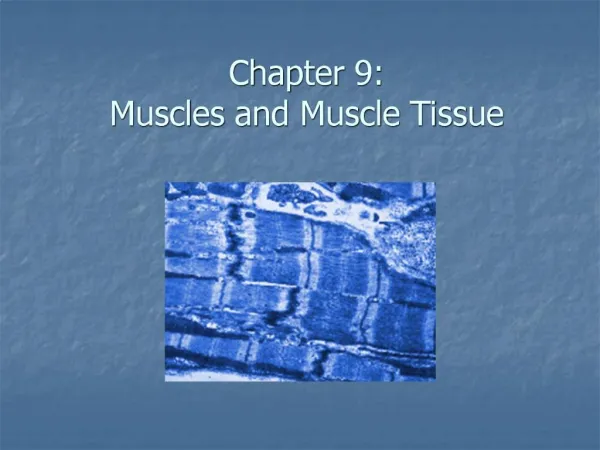
Muscles and Muscle Tissue. 8. Part A. Muscle Overview. Muscle tissue makes up nearly half the body mass. The most distinguishing functional characteristic of muscles is their ability to transform chemical energy ATP into directed mechanical energy

E N D
Muscles and Muscle Tissue 8 Part A
Muscle Overview Muscle tissue makes up nearly half the body mass. The most distinguishing functional characteristic of muscles is their ability to transform chemical energy ATP into directed mechanical energy The three types of muscle tissue are skeletal, cardiac, and smooth These types differ in structure, location, function, and means of activation
Muscle Similarities Skeletal and smooth muscle cells are elongated and are called muscle fibers Muscle contraction depends on two kinds of myofilaments – actin and myosin Muscle terminology is similar Sarcolemma – muscle plasma membrane Sarcoplasm – cytoplasm of a muscle cell Prefixes – myo, mys, and sarco all refer to muscle
Skeletal Muscle Tissue Packaged in skeletal muscles that attach to and cover the bony skeleton Has obvious stripes called striations Is controlled voluntarily (i.e., by conscious control) Contracts rapidly but tires easily Is responsible for overall body motility Is extremely adaptable and can exert forces ranging from a fraction of an ounce to over 70 pounds Key words: skeletal, striated and voluntary
Cardiac Muscle Tissue Occurs only in the heart (“blood pump”) Is striated like skeletal muscle but is not voluntary Contracts at a fairly steady rate set by the heart’s pacemaker Neural controls allow the heart to respond to changes in bodily needs Key words: cardiac, striated and involuntary
Smooth Muscle Tissue Found in the walls of hollow visceral organs, such as the stomach, urinary bladder, and respiratory passages Forces food and other substances through internal body channels It is not striated and is involuntary Key words: visceral, non striated and involuntary
Functional Characteristics of Muscle Tissue Excitability, or irritability – the ability to receive and respond to stimuli Contractility – the ability to shorten forcibly Extensibility – the ability to be stretched or extended Elasticity – the ability to recoil and resume the original resting length
Muscle Function Skeletal muscles are responsible for all locomotion Cardiac muscle is responsible for coursing the blood through the body Smooth muscle helps maintain blood pressure, and squeezes or propels substances (i.e., food, feces) through organs Muscles also maintain posture, stabilize joints, and generate heat
Skeletal Muscle Gross Anatomy Tissues: Blood vessels Nerves – branches to each fiber Connective Tissue Endomysium –wraps each fiber Perimysium –wraps fibers into fascicles Epimysium –wraps fascicles into a muscle All are continuous with each other and the tendons.
Skeletal Muscle ( Table 9.1) Figure 9.2 (a)
Skeletal Muscle: Nerve and Blood Supply Each muscle is served by one nerve, an artery, and one or more veins Each skeletal muscle fiber is supplied with a nerve ending that controls contraction Contracting fibers require continuous delivery of oxygen and nutrients via arteries Wastes must be removed via veins
Structural Organization of Skeletal Muscle Muscle (Organ) Fascicles (Bundles of fibers) Fiber (Cell) Myofibrils (densely packed contractile elements) Myofilaments (contractile proteins) Actin (thin filaments) Myosin (thick filaments)
Myofibrils Figure 9.3 (b)
Sarcomeres The smallest contractile unit of a muscle The region of a myofibril between two successive Z discs Composed of myofilaments made up of contractile proteins Myofilaments are of two types – thick and thin
Sarcomeres Figure 9.3 (c)
Myofilaments: Banding Pattern Thick filaments (composed by myosin) – extend the entire length of an A band Thin filaments (composed by actin) – extend across the I band and partway into the A band Z-disc – coin-shaped sheet of proteins (connectins) that anchors the thin filaments and connects myofibrils to one another
Myofilaments: Banding Pattern Figure 9.3 (c, d)
Ultrastructure of Myofilaments: Thick Filaments Thick filaments are composed of the protein myosin Each myosin molecule has a rodlike tail and two globular heads Tails – two interwoven, heavy polypeptide chains Heads – two smaller, light polypeptide chains called cross bridges
Ultrastructure of Myofilaments: Thick Filaments Figure 9.4 (a)(b)
Ultrastructure of Myofilaments: Thin Filaments Thin filaments are chiefly composed of the protein actin Each actin molecule is a helical polymer of globular subunits called G actin The subunits contain the active sites to which myosin heads attach during contraction Tropomyosin and troponin are regulatory subunits bound to actin
Ultrastructure of Myofilaments: Thin Filaments Figure 9.4 (c)
Arrangement of the Filaments in a Sarcomere Longitudinal section within one sarcomere Figure 9.4 (d)
Sliding Filament Model of Contraction Thin filaments slide past the thick ones so that the actin and myosin filaments overlap to a greater degree In the relaxed state, thin and thick filaments overlap only slightly Upon stimulation, myosin heads bind to actin and sliding begins
Sliding Filament Model of Contraction Each myosin head binds and detaches several times during contraction, acting like a ratchet to generate tension and propel the thin filaments to the center of the sarcomere As this event occurs throughout the sarcomeres, the muscle shortens
Skeletal Muscle Contraction In order to contract, a skeletal muscle must: Be stimulated by a nerve ending Propagate an electrical current, or action potential, along its sarcolemma Have a rise in intracellular Ca2+ levels, the final trigger for contraction
Brain Spinal Cord Motor Neuron Muscle Motor = movement Neuron = nerve cell
Motor Neuron Muscle Fiber Motor neuron releases neurotransmitter called acetylcholine (ACh). ACh causes muscle fiber to produce an electrical signal. Electrical signal causes actin & myosin to move, and this causes muscle to move.
Destruction of Acetylcholine ACh bound to ACh receptors is quickly destroyed by the enzyme acetylcholinesterase This destruction prevents continued muscle fiber contraction in the absence of additional stimuli
A Myofibril at Rest At rest” = fiber has not received message to move Actin (thin) attaches to Z-line. Myosin (thick) does not attach to actin or the Z-line. Myosin heads are bent back (cocked).
Contraction Let’s compare length of sarcomeres: Relaxed: Contracted:
Contraction in the Myofibril After contraction, muscle relaxes (back to original length). Myosin heads pick up ATP & break it apart. This cocks the head, detaching it from actin. Now ready to contract again. ATP Head is Cocked
Sequential Events of Contraction Cross bridge formation – myosin cross bridge attaches to actin filament Working (power) stroke – myosin head pivots and pulls actin filament toward M line Cross bridge detachment – ATP attaches to myosin head and the cross bridge detaches “Cocking” of the myosin head – energy from hydrolysis of ATP cocks the myosin head into the high-energy state
Contraction of Skeletal Muscle (Organ Level) Contraction of muscle fibers (cells) and muscles (organs) is similar The two types of muscle contractions are: Isometric contraction – increasing muscle tension (muscle does not shorten during contraction) Isotonic contraction – decreasing muscle length (muscle shortens during contraction)
Types of muscular contraction Isometric Muscular contraction where the tension developed occurs with no change in length Otherwise known as static contraction or position. Improves muscular strength at fixed joint angle Does not develop aerobic fitness Can be done anywhere Examples; rugby scrum, tug of war.
Types of muscle contraction Isotonic Muscles contact at speed controlled by the performer Motor unit recruitment is at the speed required for the specific sports activity. Develops aerobic and anaerobic fitness Most physical activities are isotonic Can occur in two ways: concentric and eccentric.
Contraction Concentric contraction Muscle shortens under tension Insertion moves towards origin Occurs in agonist muscle e.g. Chin-ups – use of bicep brachii in upward phase
Contraction Eccentric contraction Muscle lengthens under tension Insertion moves away from origin Occurs in antagonist muscle Only occurs if the antagonist is acting as a brake to help control the joint movement E.g. Chin-ups – use of biceps in downward phase.