Download
1 / 60
600 likes | 662 Views
Explore the relationship between blood flow and cardiac output, pressure differences in vessels, and components of total peripheral resistance in the cardiovascular system. Learn about arterial blood pressure, pulse, and the importance of resistance in maintaining blood flow. Discover how cardiovascular pressures and peripheral resistance impact circulation and overall cardiovascular health.

E N D
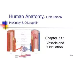
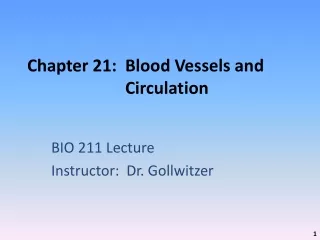
Chapter 21: Blood Vessels and Circulation BIO 211 Lecture Instructor: Dr. Gollwitzer
Today in class we will discuss: • The relationship between blood flow and • Cardiac output • Pressure and resistance • Pressure differences in various vessels in the CVS • Blood pressure , mean arterial pressure, and pulse • The basis for systolic pressure and diastolic pressure • Total peripheral resistance and its major components
Cardiovascular Physiology • Goal of cardiovascular physiology • To maintain adequate blood flow through peripheral tissues and organs • Cardiovascular system (CVS) continuously adjusted to maintain homeostasis • Contracting ventricle must produce enough tension to force open the semilunar valve and eject blood • Determined by interplay between pressure and resistance in cardiovascular network • If no resistance to blood flow, heart would not have to generate pressure to force blood through pulmonary and systemic circuits
Cardiovascular Physiology Figure 21-8
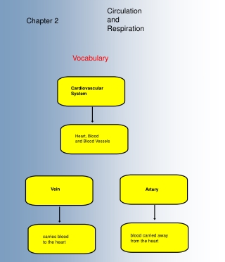
Cardiovascular Physiology • Cardiac output (CO) • Normally = blood flow • When CO goes up, blood flow through the capillary beds goes up; and vice versa • Blood (arterial) pressure (BP) • Responsible for maintaining blood flow within capillaries • Peripheral resistance controls: • Blood flow • Capillary pressure • Drives exchange via diffusion and osmosis between blood and interstitial fluid • Venous pressure • Due to venoconstriction • Aided by valves, skeletal muscle contraction • Venous return • Brings blood back to heart
Cardiovascular Pressures • General concepts • Liquids, including blood, cannot be compressed • Force exerted against an enclosed liquid (blood in CVS) generates hydrostatic pressure • If pressure gradient exists, hydrostatic pressure will push liquid from an area of higher pressure to an area of lower pressure
Cardiovascular Pressures • Pressure gradient of systemic circuit = circulatory pressure (approx 100 mm Hg) • Difference between pressure at base of ascending aorta (100 mm Hg) and entrance to R atrium (2 mm Hg) • This pressure needed to force blood through arterioles (resistance vessels) and into peripheral capillaries
Cardiovascular Pressures • 3 components • Blood pressure (BP) • = force exerted against vessel walls by blood in vessels in systemic arterial system • Ranges from 100 at heart to 35 mm Hg at start of capillary network • Capillary blood flow is directly proportional to BP • Capillary hydrostatic pressure • = Pressure in capillary beds • Declines from 35 to 18 mm Hg along length of capillary • Venous pressure • = Pressure in venous system • Pressure gradient from venules to R atrium approx. 18 mm Hg (ranges from 18 to 2 mm Hg)
Pressures in the Systemic Circuit Figure 21-10
Arterial Blood Pressure • Important because it maintains blood flow through capillary beds • Must be high enough to overcome peripheral resistance • Not stable • Rises during ventricular systole and falls during ventricular diastole • Systolic pressure (SP) = peak arterial pressure during ventricular systole • Diastolic pressure (DP) = minimum arterial pressure during diastole
Arterial Blood Pressure • Measure with sphygmomanometer • Compress brachial artery • Place stethoscope over artery, distal to compress • Inflate cuff until pressure is great enough to collapse artery and blood flow stops, pulse is eliminated • Let air out slowly • When pressure is less than SP = blood enters, pulse appears • When pressure is less than DP = pulse disappears and flow is continuous • Record by separating systolic and diastolic pressures by a slash mark (e.g., 120/80) • Normal = 120/80
Arterial Blood Pressure • Pulse • = Rhythmic pressure oscillation that accompanies each heart beat • Common site: inner wrist (radial artery pressed against radius) • Pulse pressure (PP) • = Difference between systolic and diastolic pressure (i.e., SP-DP) • Mean arterial pressure (MAP) • = Diastolic pressure + (pulse pressure/3), e.g., • If SP = 120, DP = 80 • MAP = 80 + ((120-80)/3) = 80 + 13 = 93 mm Hg
Resistance • Any force that opposes movement of fluid • Resistance of CVS • Due to friction between blood and vessel walls • Opposes movement of blood • The greater the resistance, the slower the movement of blood • For circulation to occur, pressure gradient must be great enough to overcome total peripheral resistance = resistance of the entire CVS (mostly arterial resistance b/c venous resistance so low)
Peripheral Resistance • = Resistance of the arterial system • For blood to flow into peripheral capillaries the pressure gradient must be great enough to overcome peripheral resistance • 3 sources of peripheral resistance • Vascular resistance (resistance of blood vessels) • Viscosity • Turbulence
Peripheral Resistance:Vascular Resistance • = Resistance of blood vessels • Largest component of peripheral resistance • Due to friction between blood and vessel wall • Depends on: • Vessel length • Vessel diameter
Peripheral Resistance:Vascular Resistance • Vessel length • Resistance directly proportional to length, i.e., pulmonary vs. systemic circuit • Constant in adults • Vessel diameter • Much greater effect on resistance • Effects of friction occur in zone close to vessel wall • In large diameter vessel, blood near center does not encounter resistance • In small diameter vessel, nearly all blood is slowed by friction with walls • Increases exponentially as vessel diameter decreases • ½ diameter = 16 X resistance • Mechanisms that alter diameter of arterioles provide control over peripheral resistance and blood flow • Vessel diameter varies by vasodilation (bigger) and vasoconstriction (smaller) • Most resistance occurs in arterioles (smallest diameter) = resistance vessels
Peripheral Resistance: Viscosity • = Resistance to flow caused by interactions among molecules in a liquid • Low viscosity liquids (water) flow at low pressure • Blood is 4X as viscous as water due to presence of plasma proteins and blood cells • Viscosity remains stable except in anemia, polycythemia (elevated hematocrit) and other disorders that affect hematocrit
Peripheral Resistance: Turbulence • = Swirling action disturbs smooth flow of blood • Created by: • High flow rates • Between atria and ventricles • Between ventricles and trunks • In aorta • Irregular surfaces, e.g., • Scar tissue • Atherosclerotic plaques • Sudden changes in vessel diameter, e.g., • Vasoconstriction • Slows flow and increases resistance • Does not develop in small vessels except when damaged
Vascular Pathology • Arteriosclerosis • = Thickening and toughening of arterial walls; hardening of the arteries • Complications account for half of all deaths in US • CAD (coronary artery disease) = arteriosclerosis of coronary vessels • Stroke = result of arteriosclerosis of arteries supplying brain
Vascular Pathology • Arteriosclerosis (Cont.) • 2 Forms of arteriosclerosis • Focal calcification • = Gradual degeneration of smooth muscle in tunica media and deposition of Ca2+ salts • Typically involves arteries of limbs and genitals • Rapid and severe calcification may occur as complication of diabetes mellitus • Atherosclerosis • Aka: Fatty degeneration • = Damage to endothelial lining and formation of lipid deposits (plaque) in tunica media • Most common form of arteriosclerosis
Vascular Pathology • Arteriosclerosis (Cont.) • Factors involved in development • Lipid levels; high cholesterol • High lipids in blood for an extended period of time • Monocytes begin removing them and become filled with lipid droplets (foam cells) • Attach to endothelial walls, release growth factors that stimulate smooth muscle to divide near the tunica interna, thickening the vessel wall, decreasing diameter • Other monocytes migrate resulting in a fatty mass of tissue (plaque) that projects into lumen • Because cells swollen with lipids, gaps appear in endothelial lining • Platelets appear to repair, clot forms which further restricts blood flow • High blood pressure, smoking, diabetes mellitus, obesity, stress, chronic bacterial infection, chronic inflammation
Vascular Pathology • Hypertension • = Abnormally high BP (>140/90) • Increases workload on heart, L ventricle gradually enlarges, more muscle mass • Greater O2 demand, coronary circulation can’t keep pace, eventually coronary ischemia (inadequate blood supply) • Increases stress on walls of blood vessels, accelerates development of arteriosclerosis and aneurysms • Hypotension • Abnormally low BP (SP < 90 mm Hg)
Today in class we will discuss: • The importance of maintaining adequate blood flow through the capillaries and the mechanisms involved in capillary exchange • Venous return and its role in moving blood • How autoregulatory, neural, and hormonal mechanisms compensate for a reduction in blood flow and blood pressure • How the cardiovascular system responds to hemorrhage
Capillary Exchange • As blood flows through peripheral tissues, BP forces water and solutes out of plasma, across capillary walls • Most of water is reabsorbed by capillaries • A portion (approx. 3.6 L) enters the lymphatic system and eventually re-enters bloodstream
Blood-lymph Cycle • = Continuous movement of water out of the capillaries, through peripheral walls, to lymphatics, then back to bloodstream • Has 4 important functions • Ensures plasma and interstitial fluid are in constant communication • Accelerates distribution of nutrients, hormones, dissolved gases through tissues • Transports insoluble lipids and tissue proteins that can’t cross capillary walls • Flushes bacterial toxins and chemicals into immune system tissues
Capillary Exchange • 3 processes involved in moving materials across capillary walls • Filtration • Diffusion • Reabsorption
Forces Across Capillary Walls Figure 21-13
Capillary Exchange • Filtration • Driving force is hydrostatic pressure • Forces water out of a solution from high to low pressure area (35 mm Hg in capillaries vs. 25 mm Hg in tissues) • When water is forced out of capillary walls, small solute particles travel with the water • Larger molecules and protein stay in bloodstream • Because BP drops from 35 to 18 mm Hg in capillaries, filtration occurs mostly at the arterial end
Capillary Exchange • Diffusion • = Movement of ions or molecules from high to low concentration • Does not involve pressure effects • Primary route for capillary exchange
Capillary Exchange • Capillary diffusion occurs by 5 routes • Between endothelial cells, e.g., water, ions, small organic compounds (glucose, amino acids, urea) • Through channels in cell membrane, e.g., water, Na+, K+, Ca2+, Cl- ions • At fenestrated capillaries, e.g., above and large water-soluble compounds otherwise unable to leave bloodstream • Found in hypothalamus, kidneys, endocrine organs, intestinal tract • Through endothelial cell membranes, e.g., lipids (FAs, steroids), lipid-soluble materials including gases (O2 and CO2 • In sinusoids, e.g., where plasma proteins are produced and enter bloodstream in liver
Capillary Exchange • Reabsorption • Result of osmosis = diffusion of water across membrane toward higher solute concentration, e.g, blood + plasma proteins • From arterial to venous ends of capillaries, rates of filtration and reabsorption change at about 25 mm Hg • Filtration higher at beginning and reabsorption higher at end • Of the 24 L of fluid that moves out of capillaries every day, 85% is reabsorbed • Remainder enters lymphatic vessels and eventually venous system
Capillary Exchange • Summary • Hydrostatic pressure • Forces water and solutes out of capillaries • Into interstitial fluid • At arterial end of capillary • Osmotic pressure • Pulls water and solutes into capillaries • Out of interstitial fluid • At venous end of capillary
Edema • = Abnormal accumulation of interstitial fluid (ECF) • Filtration out > reabsorption in • Results from a disturbance between hydrostatic and osmotic forces at capillary level • Causes • Damage to capillary, increase BP from heart problems, blockage of lymphatic vessels, kidney failure
Venous Pressure and Venous Return • BP very low at venules (18 mm Hg), but they provide very little resistance • As blood approaches heart • Veins become larger • Resistance drops even more • Velocity of blood increases • When individual stands, venous blood entering IVC must overcome gravity
Venous Pressure and Venous Return • 2 factors help venous blood overcome gravity • Muscular compression • Muscular contraction near vein push blood toward heart because of 1-way valves • Is why standing still for long time results in little blood flow to the brain and person faints • Respiratory pump • When inhale: • Thoracic cavity expands • Pressure in pleural cavities drops • Pulls air into lungs • Also pulls blood into IVC and R atrium from smaller veins in abdominal cavity • When exhale: • Pressure in pleural cavities rises • Pushes blood into R atrium • Important during heavy exercise
Cardiovascular Regulation • 3 Regulatory mechanisms control cardiovascular function, i.e, CO and BP • Autoregulation • Local factors at tissue level cause immediate, localized adjustments • Neural mechanisms • Respond quickly to changes at specific sitesflex control • Endocrine mechanisms • Direct long-term changes
Cardiovascular Responses Figure 21-13, 8th edition
Cardiovascular Regulation:Autoregulation • Local factors change pattern of blood flow in capillary bed in response to chemical changes in interstitial fluid • Affect precapillary sphincters • Local vasodilators – dilate sphincters • Accelerate blood flow through tissues brings O2, other nutrients to restore homeostasis • e.g., low O2 or high CO2, lactic or other acid, NO, high K+ or H+, histamine, high temperatures • Local vasoconstrictors – constrict sphincters • e.g., compounds produced by platelets and damaged tissues (antihemorrhage prostaglandins and thromboxanes from platelets and WBCs) • Cause immediate, localized hemostatic adjustments • If this fails, then neural and endocrine factors activated
Cardiovascular Regulation:Neural Mechanisms • Reflexes regulated through negative feedback loop • 2 types of reflex control • Baroreceptor reflexes • Chemoreceptor reflexes
Cardiovascular Regulation • Special cardiovascular receptors monitor conditions • Baroreceptors • Monitor and respond to stretch in blood vessels and atrium • Chemoreceptors • Monitor composition of arterial blood and CSF • Respond to changes in CO2, O2, or pH in blood • Found near carotid sinus (carotid bodies) and aortic arch (aortic bodies) • Receptors trigger neural reflex arcs to neurons in cardiovascular centers in medulla oblongata
Cardiovascular Regulation:Neural Mechanisms • Baroreceptor reflexes • Stretch receptors respond to changes in BP • Found in vessel walls and heart (carotid sinuses, R atrium, aortic sinuses) • Controlled by ANS • When BP increases, CV centers • Dec HR • Cause peripheral vasodilation dec BP • When BP decreases, CV centers • Inc HR and stroke volume • Cause peripheral vasoconstriction inc BP • Maintains normal/adequate arterial pressure
Baroreceptor Reflexes Figure 21-14, 8th edition
Cardiovascular Regulation:Neural Mechanisms • Chemoreceptor reflexes • Receptors respond to changes in O2, CO2 levels and pH in blood and CSF • Found in aortic bodies, carotid bodies, and medulla oblongata • Lead to increased HR and BP by sympathetic activation or decreased by parasympathetic activation
Chemoreceptor Reflexes Figure 21–15
Cardiovascular Regulation:Endocrine Mechanisms • Short-term regulation • Of cardiac output and peripheral vasoconstriction (resistance) • With E and NE • Long-term regulation • Of BP and/or volume • With: • ADH – increases BP and volume • Angiotensin II – increases BP and volume • Erythropoietin – increases volume • Atrial natriuretic peptide (ANP) - decreases volume
Cardiovascular Regulation:Endocrine Mechanisms • ADH • Released from post pit in response to: • Low blood volume • High plasma Na+ • Angiotensin II • Immediate result is vasoconstriction that increases BP • Stimulates conservation of water at kidneys which prevents further drop in blood volume