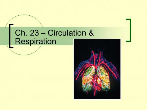
Circulation and Respiration
800 likes | 1.06k Views
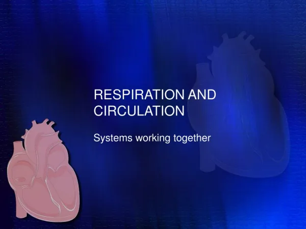
Circulation and Respiration. Chapter 22. The Circulatory System. Works with other organ systems Maintains volume, solute concentration and temperature of interstitial fluid Interstitial fluid and blood are body’s internal environment. Blood Circulation. Blood flows through blood vessels

Circulation and Respiration
E N D
Presentation Transcript
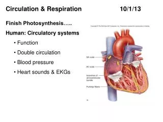
Circulation and Respiration Chapter 22
The Circulatory System • Works with other organ systems • Maintains volume, solute concentration and temperature of interstitial fluid • Interstitial fluid and blood are body’s internal environment
Blood Circulation • Blood flows through blood vessels • Heart generates force to keep blood moving • Closed system • Blood is confined to vessels and heart • Open system • Blood mingles with fluid in tissues
Open and Closed Systems aorta heart Fig. 22-1a, p.361
Open and Closed Systems pump spaces or cavities in body tissues Fig. 22-1b, p.361
Open and Closed Systems dorsal blood vessel gut cavity two of five hearts ventral blood vessels Fig. 22-1c, p.361
Open and Closed Systems pump large-diameter blood vessels (rapid flow) large-diameter blood vessels (rapid flow) small-diameter blood vessels (leisurely flow in diffusion zone) Fig. 22-1d, p.361
Blood Flow and Gas Exchange • Rate of blood flow varies with diameter of blood vessels • Slowest flow in smallest vessels, the capillaries • Gases are exchanged between blood and interstitial fluid across capillary walls
Vertebrate Circulatory Systems • Fish • Two-chambered heart, one circuit • Amphibians • Three-chambered heart, two partially separate circuits • Birds and mammals • Four-chambered heart, two entirely separate circuits
Vertebrate Circulatory Systems capillary beds of gills heart rest of body a In fishes, a two-chambered heart (atrium, ventricle) pumps blood in one circuit. Blood picks up oxygen in gills, delivers it to rest of body. Oxygen-poor blood flows back to heart. Fig. 22-2a, p.362
Vertebrate Circulatory Systems lungs right atrium left atrium heart rest of body b In amphibians, a heart pumps blood through two partially separate circuits. Blood flows to lungs, picks up oxygen, returns to heart. But it mixes with oxygen-poor blood still in the heart, flows to rest of body, returns to heart. Fig. 22-2b, p.362
Vertebrate Circulatory Systems lungs right atrium left atrium right ventricle left ventricle rest of body c In birds and mammals, the heart is fully partitioned into two halves. Blood circulates in two circuits: from the heart’s right half to lungs and back, then from the heart’s left half to oxygen-requiring tissues and back. Fig. 22-2c, p.362
Double Circuits • In birds and mammals • Right half of heart • Pulmonary circuit • Heart to lungs and return • Left half of heart • Systemic circuit • Heart to body tissues and return
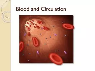
Functions of Blood • Transports oxygen and nutrients to cells • Carries carbon dioxide and wastes away from cells • Helps stabilize internal pH • Carries infection-fighting cells • Helps equalize temperature
Components of Blood • Plasma • Water • Proteins • Dissolved materials • Cells • Red blood cells • White blood cells • Platelets
Components of Blood Components Relative Amounts Plasma Portion (50%–60% of total volume): 91%–92% of plasma volume 1. Water 2. Plasma proteins (albumin, globulins, fibrinogen, etc. 7%–8% 3. Ions, sugars, lipids, amino acids, hormones, vitamins, dissolved gases 1%–2% Cellular Portion (40%–50% of total volume): 4,800,000–5,400,000 per microliter 1. Red blood cells 2. White blood cells: Neutrophils Lymphocytes Monocytes (macrophages) Eosinophils Basophils 3,000–6,750 1,000–2,700 150–720 100–360 25–90 3. Platelets 250,000–300,000 Fig. 22-3b, p.363
Blood Cell Development • Stem cells in bone marrow produce blood cells and platelets • Body continually replaces blood cells
Blood Cell Development white blood cell red blood cell platelets Fig. 22-3a, p.363
Erythrocytes (Red Cells) • Most numerous cells in blood • Transport oxygen and carbon dioxide • Colored red by oxygen-binding pigment (hemoglobin) • Have no nucleus when mature
Leukocytes (White Cells) • Function in housekeeping and defense • Cell types Basophils Dendritic cells Eosinophils B cells Neutrophils T cells Macrophages
Platelets • Membrane-bound cell fragments • Derived from megakaryocytes, which arise from stem cells • Release substances that initiate blood clotting
Human Heart Is a Double Pump • Partition separates heart into left and right sides • Each pumps blood through a different circuit
Pulmonary Circuit Heart to lungs Oxygenates blood right pulmonary artery left pulmonary artery capillary bed of left lung capillary bed of right lung pulmonary trunk (to systemic circuit) (from systemic circuit) pulmonary veins heart lungs
Systemic Circuit capillary beds of head and upper extremities aorta (to pulmonary circuit) (from pulmonary circuit) Starts at aorta Carries oxygenated blood to body tissues heart capillary beds of other organs in thoracic cavity capillary bed of liver capillary beds of intestines capillary beds of other abdominalorgans and lower extremities
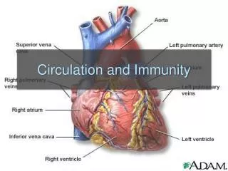
Major Vessels carotid arteries jugular veins ascending aorta superior vena cava pulmonary arteries pulmonary veins coronary arteries hepatic portal vein brachial artery renal artery renal vein inferior vena cava abdominal aorta iliac arteries iliac veins femoral artery femoral vein
Four Chambers • Each side has two chambers • Upper atrium • Lower ventricle • Valves between atria and ventricles
Major Vessels Heart Anatomy arch of aorta superior vena cava trunk of pulmonary arteries left semilunar valve right semilunar valve left pulmonary veins right pulmonary veins left atrium right atrium left AV valve right AV valve right ventricle left ventricle endothelium and connective tissue inferior vena cava inner layer of pericardium septum heart’s apex myocardium
Cardiac Cycle Diastole (mid to late). Ventricles fill, atria contract. Ventricular systole (atria are still in diastole). Ventricles eject. Diastole (early). Both chambers relax.
Conduction and Contraction • SA node in right atrium is pacemaker • Electrical signals cause contraction of atria • Signal flows to AV node and down septum to ventricles SA node
Blood Vessels • Arteries: carry blood away from heart • Arterioles: diameter is adjusted to regulate blood flow • Capillaries: diffusion occurs across thin walls
Blood Pressure • Highest in arteries, lowest in veins • Usually measured in the brachial artery • Systolic pressure is peak pressure • Ventricular contraction • Diastolic pressure is the lowest pressure • Ventricular relaxation
Resistance • Adjusted at arterioles • Vasodilation • Increases vessel diameter • Lowers blood pressure • Vasoconstriction • Decreases vessel diameter • Increases blood pressure
Distribution lungs 100% heart’s right half heart’s left half 6% liver 21% digestive tract 20% kidneys 15% skeletal muscle 13% brain 9% skin 5% bone 3% cardiac muscle 8% all other regions Fig. 22-10, p.367
Capillary Beds • Diffusion zone; site of exchange between blood and interstitial fluid • Capillary wall is one cell thick • Flow is slow; allows gases to diffuse across membranes of blood cells and across endothelium
Bulk Flow in Capillary Bed blood to venule outward-directed bulk flow inward-directed osmotic movement blood from arteriole cells of tissue
Net Bulk Flow • Normally, ultrafiltration only slightly exceeds reabsorption • Fluid enters interstitial fluid and returned to blood via the lymphatic system • High blood pressure causes excessive ultrafiltration and results in edema
The Venous System • Blood flows from capillaries to venules to veins • Veins are large-diameter vessels with some smooth muscle in wall
Vein Function • Valves in veins prevent blood from flowing backward
Vein Function valve closed blood flow to heart valve open valve closed venous valve valve closed Fig. 22-13, p.369
Hemostasis • Processes that stop blood loss and repair vessels • Blood vessel spasm • Platelet plug formation • Blood coagulation • Clotting
Clotting Mechanism • Prothrombin is converted to thrombin • Fibrinogen is converted to fibrin • Fibrin forms net that entangles cells and platelets
Hypertension • Blood pressure above 140/90 • Tends to be genetic • May also be influenced by diet • Contributes to atherosclerosis • “Silent killer”, few outward signs
Atherosclerosis • Arteries thicken, lose elasticity • Fill up with cholesterol and lipids • High LDL increases risk
wall of artery, cross-section unobstructed lumen of normal artery Fig. 22-15a, p.370
atherosclerotic plaque blood clot sticking to plaque narrowed lumen Fig. 22-15b, p.370
Coronary Artery Disease • Atherosclerosis in arteries of heart • Causes heart attacks
Coronary Artery Disease coronary artery aorta coronary artery blockage location of a shunt made of a section taken from one of the patient’s other blood vessels Fig. 22-16, p.371
Risk Factors Smoking Genetics High cholesterol High blood pressure Obesity Diabetes Age Gender
Respiration • Respiration • Physiological process by which oxygen moves into an animal’s internal environment and carbon dioxide moves out • Aerobic respiration • Cellular process, produces ATP • Oxygen is used • Carbon dioxide is produced