Download
1 / 11
110 likes | 144 Views
This study delves into abnormal zonular ligament morphology in dogs with lens displacement, particularly focused on terrier breeds. Using light microscopy, it aims to identify abnormal patterns in zonular ligament protein and potential breed correlations. The findings suggest a link between zonular ligament dysplasia, collagenization, and age-related changes.

E N D
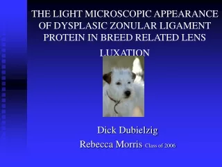
THE LIGHT MICROSCOPIC APPEARANCE OF DYSPLASIC ZONULAR LIGAMENT PROTEIN IN BREED RELATED LENS LUXATION Dick Dubielzig Rebecca Morris Class of 2006
Lens displacement is a common cause of secondary glaucoma in the dog • Lens displacements particularly common in terrier breeds • Abnormal zonular ligament morphology SEM, PAS in Tibetan Terriers, Curtis et.al. J.Comp.Path., 1983 • Lack of description of nature of abnormality using standard histopathlogy
Study Design • Hypothesis: determine whether LM can detect abnormal patterns in zonular ligament protein, any breed correlation • Search of COPLOW database for glaucoma caused by lens displacement • Selection criteria • Exclusions • Evaluation • From records: signalment and laterality • Histopathology: zonular ligament morphology, other morphological features
The normal make-up of the zonule • Fibrillin 1 & Fibrillin2 • Glycoprotein sub-units • Strung together as a “string of beads” • Surrounded by microfibril associated glycoprotein • Fibrillin also found as a component of elastin
Zonular protein classification • Normal • Abnormal • Zonular ligament dysplasia • Thick lamellar protein sheet tightly adherent to nonpigmented ciliary body epithelium • Collagenization • Filamentous, not tightly adherent
StainingCharacteristics Normal zonular protein Zonular ligament dysplasia Collagenization PAS Trichrome Elastin Stain
Results *includes 4 Shar-Peis
Average Age • Normal Zonule Profile: 10.7 years • Collagenization: 8.9 years • Zonular Ligament Dysplasia: 5.2 years
Laterality • OS: 39 • OD: 28 • Unknown: 5 • 9 dogs had both globes involved and 8 of these had zonular ligament dysplasia
Conclusions • LM is useful in detecting abnormal patterns in zonular ligament protein • “Normal” zonular ligament with glaucoma • Secondary to trauma or other disease process? • Zonular ligament dysplasia • Terriers and Shar-Peis, young, bilateral, distinct morphology • Collagenization • No breed predilection, older, possibly secondary to disease process