Download
1 / 79
790 likes | 878 Views
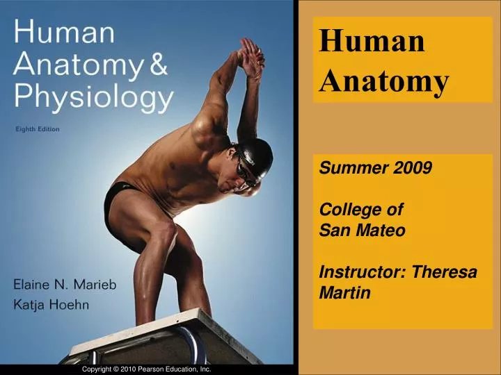
Human Anatomy. Summer 2009 College of San Mateo Instructor: Theresa Martin. Student Learning Objectives. Identify the structures of the body by systems. Relate the structure to the function of anatomic structures.

E N D
Human Anatomy Summer 2009College of San MateoInstructor: Theresa Martin
Student Learning Objectives • Identify the structures of the body by systems. • Relate the structure to the function of anatomic structures. • Manipulate cadaver dissections and other lab specimens to understand structural relationships in the body. • Learn the aspects of normal functioning in order to relate to clinical issues.
The Language of Anatomy • Originally from Latin and Greek • Word roots have specific meanings • Osteo- • Cyte-
Principle of Complementarity • Anatomy and physiology are inseparable. • Every structure has a function • What a structure can do depends on its specific form
1 The Human Body: An Orientation
Levels of Body Organization • Chemicals • Cells • Tissues • Organs • Organ Systems • Organism
Organelle Atoms Molecule Smooth muscle cell Cellular levelCells are made up ofmolecules. 2 Chemical levelAtoms combine to form molecules. 1 Smooth muscle tissue Cardiovascularsystem Tissue levelTissues consist of similartypes of cells. 3 Heart Bloodvessels Blood vessel (organ) Smooth muscle tissue Connective tissue Epithelialtissue Organ levelOrgans are made up of different typesof tissues. 4 Organismal levelThe human organism is made upof many organ systems. Organ system levelOrgan systems consist of differentorgans that work together closely. 6 5 Figure 1.1
Atoms Molecule Chemical levelAtoms combine to form molecules. 1 Figure 1.1, step 1
Organelle Atoms Molecule Smooth muscle cell Cellular levelCells are made up ofmolecules. 2 Chemical levelAtoms combine to form molecules. 1 Figure 1.1, step 2
Organelle Atoms Molecule Smooth muscle cell Cellular levelCells are made up ofmolecules. 2 Chemical levelAtoms combine to form molecules. 1 Smooth muscle tissue Tissue levelTissues consist of similartypes of cells. 3 Figure 1.1, step 3
Organelle Atoms Molecule Smooth muscle cell Cellular levelCells are made up ofmolecules. 2 Chemical levelAtoms combine to form molecules. 1 Smooth muscle tissue Tissue levelTissues consist of similartypes of cells. 3 Blood vessel (organ) Smooth muscle tissue Connective tissue Epithelialtissue Organ levelOrgans are made up of different typesof tissues. 4 Figure 1.1, step 4
Organelle Atoms Molecule Smooth muscle cell Cellular levelCells are made up ofmolecules. 2 Chemical levelAtoms combine to form molecules. 1 Smooth muscle tissue Cardiovascularsystem Tissue levelTissues consist of similartypes of cells. 3 Heart Bloodvessels Blood vessel (organ) Smooth muscle tissue Connective tissue Epithelialtissue Organ levelOrgans are made up of different typesof tissues. 4 Organ system levelOrgan systems consist of differentorgans that work together closely. 5 Figure 1.1, step 5
Organelle Atoms Molecule Smooth muscle cell Cellular levelCells are made up ofmolecules. 2 Chemical levelAtoms combine to form molecules. 1 Smooth muscle tissue Cardiovascularsystem Tissue levelTissues consist of similartypes of cells. 3 Heart Bloodvessels Blood vessel (organ) Smooth muscle tissue Connective tissue Epithelialtissue Organ levelOrgans are made up of different typesof tissues. 4 Organismal levelThe human organism is made upof many organ systems. Organ system levelOrgan systems consist of differentorgans that work together closely. 6 5 Figure 1.1, step 6
Homework What are the organ systems of the human body? What organs are in each system? What does each organ system do?
Organ Systems Interrelationships • All cells depend on organ systems to meet their survival needs • Organ systems work together to perform necessary life functions
Digestive system Takes in nutrients, breaks them down, and eliminates unabsorbed matter (feces) Respiratory system Takes in oxygen and eliminates carbon dioxide Food O2 CO2 Cardiovascular system Via the blood, distributes oxygen and nutrients to all body cells and delivers wastes and carbon dioxide to disposal organs Blood CO2 O2 Urinary system Eliminates nitrogenous wastes and excess ions Heart Nutrients Interstitial fluid Nutrients and wastes pass between blood and cells via the interstitial fluid Integumentary system Protects the body as a whole from the external environment Feces Urine Figure 1.2
3 Cells: The Living Units
Cell Theory • The cell is the smallest unit of life
Cell Diversity • Over 200 different types of human cells • Types differ in size, shape, intracellular components, and functions
Erythrocytes Fibroblasts Epithelial cells (a) Cells that connect body parts, form linings, or transport gases Nerve cell Skeletal Muscle cell (e) Cell that gathers information and control body functions Smooth muscle cells (b) Cells that move organs and body parts Sperm Macrophage (f) Cell of reproduction Fat cell (c) Cell that storesnutrients (d) Cell that fights disease Figure 3.1
Generalized Cell • All cells have some common structures and functions • Human cells have three basic parts: • Plasma membrane—flexible outer boundary • Cytoplasm—intracellular fluid containing organelles • Nucleus—control center
Nuclear envelope Chromatin Nucleolus Nucleus Smooth endoplasmic reticulum Plasma membrane Mitochondrion Cytosol Lysosome Centrioles Centrosome matrix Rough endoplasmic reticulum Ribosomes Golgi apparatus Secretion being released from cell by exocytosis Cytoskeletal elements • Microtubule • Intermediate filaments Peroxisome Figure 3.2
Plasma Membrane • Bilayer of lipids and proteins in a constantly changing fluid mosaic • Separates intracellular fluid (ICF) from extracellular fluid (ECF) • Interstitial fluid (IF) = ECF that surrounds cells
Extracellular fluid (watery environment) Cholesterol Polar head of phospholipid molecule Glycolipid Glycoprotein Carbohydrate of glycocalyx Outward- facing layer of phospholipids Integral proteins Filament of cytoskeleton Peripheral proteins Bimolecular lipid layer containing proteins Inward-facing layer of phospholipids Nonpolar tail of phospholipid molecule Cytoplasm (watery environment) Figure 3.3
Membrane Lipids • 75% phospholipids (lipid bilayer) • 5% glycolipids • 20% cholesterol
Extracellular fluid (watery environment) Cholesterol Polar head of phospholipid molecule Glycolipid Glycoprotein Carbohydrate of glycocalyx Outward- facing layer of phospholipids Integral proteins Filament of cytoskeleton Peripheral proteins Bimolecular lipid layer containing proteins Inward-facing layer of phospholipids Nonpolar tail of phospholipid molecule Cytoplasm (watery environment) Figure 3.3
Membrane Proteins • Integral proteins • Firmly inserted into the membrane (most are transmembrane) • Functions: • Transport proteins (channels and carriers), enzymes, or receptors
Membrane Proteins • Peripheral proteins • Loosely attached to integral proteins • Include filaments on intracellular surface and glycoproteins on extracellular surface • Functions: • Enzymes, motor proteins, cell-to-cell links, provide support on intracellular surface, and form part of glycocalyx PLAY Animation: Structural Proteins PLAY Animation: Receptor Proteins
Extracellular fluid (watery environment) Cholesterol Polar head of phospholipid molecule Glycolipid Glycoprotein Carbohydrate of glycocalyx Outward- facing layer of phospholipids Integral proteins Filament of cytoskeleton Peripheral proteins Bimolecular lipid layer containing proteins Inward-facing layer of phospholipids Nonpolar tail of phospholipid molecule Cytoplasm (watery environment) Figure 3.3
Functions of Membrane Proteins • Transport • Receptors for signal transduction • Attachment to cytoskeleton and extracellular matrix
(a) Transport A protein (left) that spans the membrane may provide a hydrophilic channel across the membrane that is selective for a particular solute. Some transport proteins (right) hydrolyze ATP as an energy source to actively pump substances across the membrane. Figure 3.4a
(b) Receptors for signal transduction Signal A membrane protein exposed to the outside of the cell may have a binding site with a specific shape that fits the shape of a chemical messenger, such as a hormone. The external signal may cause a change in shape in the protein that initiates a chain of chemical reactions in the cell. Receptor Figure 3.4b
(c) Attachment to the cytoskeleton and extracellular matrix (ECM) Elements of the cytoskeleton (cell’s internal supports) and the extracellular matrix (fibers and other substances outside the cell) may be anchored to membrane proteins, which help maintain cell shape and fix the location of certain membrane proteins. Others play a role in cell movement or bind adjacent cells together. Figure 3.4c
Functions of Membrane Proteins • Enzymatic activity • Intercellular joining • Cell-cell recognition
(d) Enzymatic activity Enzymes A protein built into the membrane may be an enzyme with its active site exposed to substances in the adjacent solution. In some cases, several enzymes in a membrane act as a team that catalyzes sequential steps of a metabolic pathway as indicated (left to right) here. Figure 3.4d
(e) Intercellular joining Membrane proteins of adjacent cells may be hooked together in various kinds of intercellular junctions. Some membrane proteins (CAMs) of this group provide temporary binding sites that guide cell migration and other cell-to-cell interactions. CAMs Figure 3.4e
(f) Cell-cell recognition Some glycoproteins (proteins bonded to short chains of sugars) serve as identification tags that are specifically recognized by other cells. Glycoprotein Figure 3.4f
Membrane Junctions • Three types: • Tight junction • Desmosome • Gap junction
Microvilli Plasma membranes of adjacent cells Intercellular space Basement membrane Interlocking junctional proteins Intercellular space (a) Tight junctions:Impermeable junctions prevent molecules from passing through the intercellular space. Figure 3.5a
Membrane Junctions: Tight Junctions • Prevent fluids and most molecules from moving between cells • Where might these be useful in the body?
Microvilli Plasma membranes of adjacent cells Intercellular space Basement membrane Intercellular space Plaque Intermediate filament (keratin) Linker glycoproteins (cadherins) (b) Desmosomes: Anchoring junctions bind adjacent cells together and help form an internal tension-reducing network of fibers. Figure 3.5b
Membrane Junctions: Desmosomes • “Rivets” or “spot-welds” that anchor cells together • Where might these be useful in the body?
Plasma membranes of adjacent cells Microvilli Intercellular space Basement membrane Intercellular space Channel between cells (connexon) (c) Gap junctions: Communicating junctions allow ions and small mole- cules to pass from one cell to the next for intercellular communication. Figure 3.5c
Membrane Junctions: Gap Junctions • Transmembrane proteins form pores that allow small molecules to pass from cell to cell • For spread of ions between cardiac or smooth muscle cells
Membrane Transport • Plasma membranes are selectively permeable • Some molecules easily pass through the membrane; others do not
Types of Membrane Transport • Passive processes • No cellular energy (ATP) required • Substance moves down its concentration gradient • Active processes • Energy (ATP) required • Occurs only in living cell membranes
Passive Processes • What determines whether or not a substance can passively permeate a membrane? • Lipid solubility of substance • Channels of appropriate size • Carrier proteins PLAY Animation: Membrane Permeability
Passive Processes • Simple diffusion • Carrier-mediated facilitated diffusion • Channel-mediated facilitated diffusion • Osmosis
Passive Processes: Simple Diffusion • Nonpolar lipid-soluble (hydrophobic) substances diffuse directly through the phospholipid bilayer PLAY Animation: Diffusion
Extracellular fluid Lipid- soluble solutes Cytoplasm (a) Simple diffusion of fat-soluble molecules directly through the phospholipid bilayer Figure 3.7a