Download

1 / 21
210 likes | 338 Views
Learn about cardiac muscle types, anatomy, and contraction steps. Explore the unique features of cardiac muscle cells and their contractile activity. Discover the excitation-contraction coupling process in cardiac muscle.

E N D
CARDIAC MUSCLE OBJECTIVES At the end of this lecture you should be able to know types of cardiac muscle and its anatomical location. • Arrangement of myofilaments and sarcoplasmic reticulum • Steps involved in Contraction • Functional differences
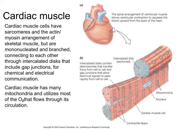
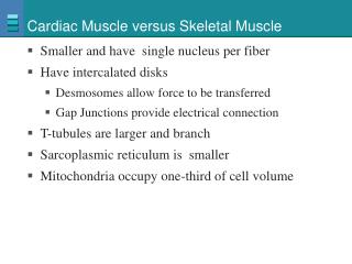
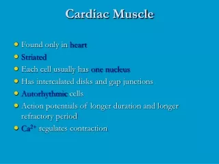
CARDIAC MUSCLES • Cardiac muscle is found only in heart. • Shape = Rectangular shape.Short branched cells • Diameter = 20um Length = 100 um • Single nucleus • Regeneration of cardiac muscle fiber is controversial.Recent studies suggest that cardiac muscle cells have mild proliferative property.
Cardiac muscle • Thick myosin filaments. • Thin actin filaments (Troponin present) • Striated appearance. • T-Tubule system associated with calcium loaded sarcoplasmicretculum. • Contraction occurs by sliding filament mechanism.
Intercalated Disks • Structures between adjacent cells. • Myofibrils attached to desmosomes. • Gap junctions present.
Input influencing Cardiac muscle contractile activity. 1. Sinoatrial node (SA node-Pacemaker) Location=Superior posterolateral wall of right atrium. Hormones. Neurotransmitters
Sources of Cytosolic Calcium • L-Type calcium channels (modified DPR) (L= long lasting current )—Cardiac muscle cannot under go tetanic contractions. • ECF calcium 1.Depolarization of plasma membrane. 2.Slight Increase in cytosolic Ca 3.Triggers release of Ca from SR. • Rynaodine receptors (Ca channels)
Thin filament activation • Cross- bridge cycling • Force generation • Same as SKELETAL MUSCLE
Contraction ends • Cytosolic Ca conc is restored Ca-ATPase pumps Na/Ca countertranspoters
Contraction ends • Cytosolic Ca conc is restored. Ca-ATPase activity Na/Ca countertranspoters