Gas Exchange Surfaces
490 likes | 547 Views
This text explores the process of gas exchange in respiratory systems and the different surfaces involved. It discusses ventilation, external respiration, and internal respiration, as well as the importance of vascularization and respiratory pigments. The difficulties of obtaining oxygen in water environments compared to air are also explained, along with examples of gas exchange in aquatic and terrestrial animals. The text covers the anatomy of gills in bony fishes, respiration in terrestrial environments, tracheae in insects, and lungs of vertebrates.

Gas Exchange Surfaces
E N D
Presentation Transcript
Gas Exchange Surfaces • Respiration: • The events associated with gas exchange between the cells and the external environment • Consists of these steps: • Ventilation = inspiration (air in) & expiration (air out) • External Respiration = gas exchange between external environment & the blood within respiration surfaces. Blood then transports oxygen to the tissues. • Internal Respiration = gas exchange between blood & tissue fluid. Cells exchange gases with tissue fluid. Blood transports carbon dioxide back to respiratory surfaces.
Gas Exchange Surfaces • For diffusion to be effective, gas-exchange tissues must be: • Moist because gases must be in solution • Thin to allow for rapid diffusion • Relatively large in relation to size of body to ensure that cells get oxygen in a timely fashion Relatively small, and flat, animals don’t need a specialized respiration system: Planaria - flat, 2-dimensional body allows surface of animal to be gas-exchange surface Larger, more 3-dimensional animals need specialized gas-exchange surfaces such as gills or lungs.
Gas Exchange Surfaces • Effectiveness of diffusion is enhanced by vascularization: • • Gas-exchange surfaces are usually associated with capillary beds so that oxygen and carbon dioxide can be exchanged efficiently. • Delivery of oxygen to cells is promoted by respiratory pigments such as hemoglobin that can pick up the oxygen and carry it.
Gas Exchange in Water Environments • I. Difficulties obtaining oxygen in water compared to air: • A. Water contains only a fraction of the oxygen that would be present in the same volume of air • 1. Oxygen has low solubility in water • 0.004% in seawater; 21% in air. • B. Diffusion of oxygen in water is thousands of times slower than in air • C. Water is more dense than air • 1. Use more energy to respire than do land animals. • •Fish use up to 25% of energy output to respire while terrestrial animals only use 1-2% of their energy output.
Gas Exchange in Water Environments • II. Small, simple multicellular animals: • A. Planaria, hydra • 1. Gastrovascular cavity helps put cells in contact with oxygenated water • B. Aquatic worms • 1. Use skin that is supplied with blood vessels
Gas Exchange in Aquatic Environments • III. Larger aquatic animals like bony fishes •Often have gills: • Outward extensions of pharynx; said to be evaginated • Have finely subdivided surfaces with a huge total surface area • Contain a rich supply of blood vessels (vascularized) • Ventilation is brought about by combined action of the mouth drawing water in and gill covers (operculum) forcing water out of head area
Countercurrent Exchange System • Countercurrent Exchange System in Fish Gills: •In lamella of gills: • Blood flows in the direction opposite to the movement of water across the gills. • This maximizes the amount of oxygen picked up by the blood: - As the blood gains oxygen, it always encounters water having an even higher oxygen content. - No equilibrium is ever reached. 80-90% of dissolved oxygen is extracted. If flow of blood was concurrent to water (same direction) an equilibrium point would occur and less oxygen would be transferred into blood.
Respiration in Terrestrial Environments: • Air has much more available oxygen than water but it is a drying environment. Thus, terrestrial animals tend to have invaginated respiratory systems to protect them from too much water loss. • - Exception is earthworms which breathe through their skin but must keep body surface moist by secreting mucus, etc. • - Earthworms also do their best to remain in damp soil during the daytime.
Land Environments:Tracheae • Insects and other terrestrial arthropods • Respiratory system consists of branched air tubes called tracheae • Oxygen enters tracheae at spiracles, valvelike openings on each side of the body. • Tracheae branch & branch until they end in tiny channels, the tracheoles, that are in direct contact with body cells. • Very efficient system for delivering oxygen to cells that does NOT involve any respiratory pigments or circulatory system.
Land Environments:Lungs of Vertebrates • Terrestrial vertebrates have evolved lungs • Vascular outgrowths from lower pharyngeal region • Lungs of amphibians • Possess a short tracheae which divides into two bronchi that open into lungs • Many also breathe to some extent through skin • Reptiles • Inner lining of lungs is more finely divided in reptiles than in amphibians • Lungs of birds and mammals are elaborately subdivided • All terrestrial vertebrates, except birds, use a tidal ventilation system • Air moves in and out by the same route. Thus, fresh incoming air is mixed with some left-over stale air.
Ventilation in Frogs Amphibians use both negative &positive pressure to ventilate their lungs: 1. Negative pressure: With mouth closed but nostrils open, the floor of the mouth is lowered. The lower air pressure will cause air to rush into their mouth cavity. 2. Positive pressure: With mouth & nostrils shut, floor of mouth rises & pushes air into the lungs.
Ventilation inTerrestrial Vertebrates • Inspiration in mammals (Inhalation) • Create negative pressure in lungs • The rib cage is elevated by intercostal muscles. • The diaphragm contracts and pushes down towards belly • Thoracic pressure decreases to less than atmospheric pressure • Atmospheric pressure forces air into the lungs • Expiration in mammals (Exhalation) • Create positive pressure in lungs • The rib cage is lowered as intercostal muscles relax • The diaphragm relaxes and rises back up towards chest • Thoracic pressure increases to more than atmospheric pressure • Forces air out the lungs
Ventilation in Birds • Birds use a one-way ventilation mechanism in lungs • How does this work? - Incoming air does not directly enter the lungs, instead it is carried by trachea to a set of posteriorair sacs. - Air is then pushed through tiny tubes called parabronchi which are surrounded by capillaries - Air ends up in anterior air sacs which expel it from body. Thus, fresh & used air never mix in the lungs of birds. • Results in a higher partial pressure of oxygen in the lungs • Oxygen uptake with each breath is greater than in other vertebrates
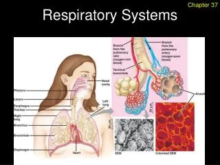
Human Respiratory System • As air moves through upper respiratory system (nostrils, nasal cavities, pharynx, larynx, trachea): • It is filtered to free it of debris (cilia help this • Warmed, and • Humidified • When air reaches lungs • It is at body temperature, and • Its humidity is 100%
Human Respiratory System • Air passes from pharynx through glottis, an opening in larynx • Larynx (voice box) contains vocal cords which allow us to produce sounds • Trachea • Permanently held open by cartilage rings • Facilitates movement of air • When food is swallowed: • The larynx rises, and • The glottis is closed by a flap of tissue called the epiglottis • Backward movement of soft palate covers the entrance of nasal passages into the pharynx
Human Respiratory System • Trachea divides and forms two primary bronchi • Bronchi enter the right and left lungs • Bronchi branch until there are a great number of tiny bronchioles. The walls of bronchioles get thinner & rings of cartilage are no longer present. • Each bronchiole terminates in an elongated space enclosed by a great number of air pockets, or sacs, called alveoli. Gas exchange occurs between air in sacs and blood in surrounding capillaries.
Control of Breathing Rate in Humans • Average person takes a breath about 14 times per minute when at rest • Respiratory control center, located in pons & medulla oblongata of the brain, can change the normal rate according to circumstances. - When a drop in pH is noted (due to increase in CO2) the control center increases rate & depth of breathing. - Normally, O2 concentration in blood has little effect on breathing rate. However, if O2 level is very low detectors in aorta & carotid arteries send an alarm to resp. control centers in brain.
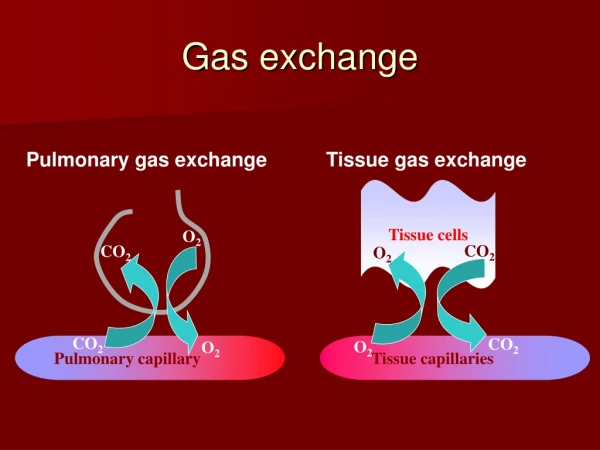
Gas Exchange and Transport • External Respiration • 1. Blood flowing into pulmonary capillaries has higher CO2 concentration than air in the alveolar air sacs. • CO2 diffuses out of pulmonary capillaries & into air sacs. 2. Blood coming into pulmonary capillaries has lower concentration of O2 than alveolar air. O2 diffuses from alveoli into capillaries.
Transport of Oxygen • Most oxygen that enters the pulmonary capillaries combines with hemoglobin in red blood cells to form oxyhemoglobin. • - Each of the 4 polypeptide chains of hemoglobin is folded around a heme (iron) group. • Iron forms a loose bond with oxygen. • •At normal partial pressure of O2 in lungs, hemoglobin is almost saturated with oxygen. • •At partial pressure of O2 in tissues, oxyhemoglobin gives up much of its oxygen during internal respiration. • - Acidity & warm temperatures promote this response in the tissues.
Transport of Carbon Dioxide • Internal Respiration • Carbon dioxide enters blood from the tissues • 1. Some carbon dioxide combines with hemoglobin to form carbaminohemoglobin • 2. Most carbon dioxide is transported in the form of bicarbonate ions (HCO3-) • - CO2 combines with water, forming carbonic acid (H2CO3) & then dissociates into H+ & HCO3-. • - Carbonic anhydrase, an enzyme, speeds up this reaction. • - H+ combines with globin part of hemoglobin (HHb) & HCO3- diffuses into plasma. This plays vital role in maintaining proper pH of blood.
Transport of Carbon Dioxide • External Respiration • 1. As blood enters the pulmonary capillaries, most of the CO2 is present in plasma as HCO3- • 2. HHb gives up the H+ it has been carrying & carbonic anhydrase speeds up this reaction: H+ + HCO3- H2CO3 H2O + CO2 3. Now free CO2 diffuses out of blood into the alveoli of lungs.
Respiration and Health • Upper Respiratory Tract Infections • Consists of nose, pharynx & larynx. Infections can spread from nasal cavities to sinuses, to middle ears & to larynx • Strep Throat • Usually starts as viral infection that becomes a secondary bacterial infection. • Caused by Streptococcus pyogenes. Can become generalized upper respiratory infection. • Symptoms: severe sore throat, high fever, white patches on dark red throat
Respiration and Health • Sinusitis • Infection of sinuses, facial cavities that drain into nasal cavities • Develops when nasal congestion blocks openings into the sinuses • Tonsillitis • Infection of tonsils, masses of lymphatic tissue. • Tonsils help to remove pathogens from pharynx • Laryngitis • Infection of larynx accompanied by hoarseness & possibly an inability to talk.
Respiration and Health • Lower Respiratory Tract Infections • Infections of trachea, bronchi, bronchioles & lungs • Acute bronchitis • Infection of primary and secondary bronchi • Usually preceded by a viral upper respiratory infection that led to secondary bacterial infection
Respiration and Health • Pneumonia • Viral, bacterial or fungal infection of the lungs in which bronchi and alveoli fill with pus & fluid • Most often preceded by influenza, the “flu”. • Can be localized in specific lobules of lungs; more lobules the more serious the infection • AIDS patients often get a rare pneumonia caused by a fungus called Pneumocystis carinii.
Respiration and Health • Pulmonary tuberculosis (TB) • Caused by tubercle bacillus, a type of bacterium. • Can test people with a simple skin test to see if they have been exposed to tuberculosis • Reaction to bacterium: 1. When the bacteria invade the lung, the cells build a protective capsule around the bacteria. This capsule is called a tubercle. 2. With a good immune system the body might kill the encapsulated bacteria 3. With a weakened immune system, like in AIDS, the bacteria can be released & hurt the body
Disorders • Pulmonary fibrosis • Fibrous connective tissue builds up in the lungs • Due to inhalation of particles such as silica, coal dust, asbestos & fiberglass. • Lungs can’t inflate properly • Asbestos also associated with cancer • Chronic bronchitis • Airways inflamed and filled with mucus • Coughing causes bronchi to undergo changes, including loss of cilia & normal cleansing action • Most frequent cause is smoking.
Disorders • Emphysema • Alveoli are distended (stretched) and walls are damaged reducing surface area available for gas exchange • Often preceded by chronic bronchitis • Elastic recoil of lungs is reduced; thus expiration is very difficult • Heart works harder to force more blood to lungs • Symptoms:breathlessness & cough, depression & irritability
Disorders • Asthma • Airways are unusually sensitive to specific irritants • When exposed to the irritants, the smooth muscles in the bronchioles undergo spasms • Irritants can be pollen, animal dander, dust, cigarette smoke, fumes & even cold air • Not curable but is treatable with inhalers that can control inflammation of bronchioles & prevent attack or stop muscle spasms during an attack
Disorders • Lung Cancer • Begins with thickening and callusing of the cells lining the airways • Loss of cilia follows; thus it is impossible to prevent dust & dirt from settling into lungs • Atypical nuclei appear in callused lining • Creates a tumor of such cells • Final step is when some cells break loose & penetrate other tissues (metastasis)