Download
1 / 73
770 likes | 1.99k Views
STRESS ECG AND STRESS ECHOCARDIOGRAPHY. Giuseppe Biondi Zoccai Division of Cardiology , University of Turin , Turin , Italy Meta-analysis and Evidence-based medicine Training in Cardiology (METCARDIO), Ospedaletti , Italy. LEARNING GOALS. Scope of the problem Stress ECG

E N D
STRESS ECG AND STRESS ECHOCARDIOGRAPHY Giuseppe Biondi Zoccai DivisionofCardiology, UniversityofTurin, Turin, Italy Meta-analysis and Evidence-based medicine Training in Cardiology(METCARDIO), Ospedaletti, Italy
LEARNING GOALS • Scope of the problem • Stress ECG • Stress echocardiography • Reconciling the evidence
LEARNING GOALS • Scope of the problem • Stress ECG • Stress echocardiography • Reconciling the evidence
DIAGNOSTIC AND PROGNOSTIC WORK-UP OF SUSPECTED CORONARY HEART DISEASE • Clinical history • Physical examination • Resting ECG • Resting echocardiography • Stress ECG • Stress echocardiography • Stress nuclear scan • Coronary CT • Coronary angiography • ….
EFFECTIVE RADIATION DOSES Picano, Am J Med 2003
CORONARY STEAL PHENOMENON Picano, Circ 1998
CORONARY STEAL PHENOMENON Picano, Circ 1998
THREE STATES OF THE SODIUM CHANNEL AND THE NORMAL SODIUM CURRENT (INa) 0 Late Na+ SodiumCurrent Na+ Na+ Peak Activated Resting Closed Inactivated out Na+ in Na+ [Na] 140 mM Na+ Na+ Na+ Na+ ~ 10mM Ca++ Ca++ in Ca++ Ca++ Na+ Ca++ Ca++ out Na+/Ca++ Exchanger Na+ Ca++
Excess Calcium: • Electrical instability • Contractile dysfunction • ECG changes ISCHEMIA INDUCED EFFECTS ON LATE INa AND INTRACELLULAR CALCIUM 0 SodiumCurrent Late Na+ Impaired Inactivation Peak Na+ Na+ out Na+ Na+ Na+ Ca++ Na+ Na+ in Na+ Na+ Na+ Na+ Ca++ Ca++ Ca++ Ca++ Ca++ Ca++ Ca++ Ca++ in Ca++ Ca++ Na+ Ca++ Ca++ Ca++ Ca++ out Na+/Ca++ Exchanger Na+ Ca++
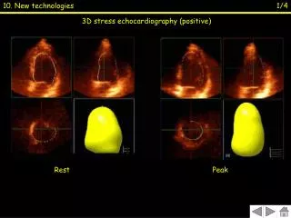
LEARNING GOALS • Scope of the problem • Stress ECG • Stress echocardiography • Reconciling the evidence
EQUIPMENT FOR STRESS TESTING • Treadmill or bicycle or steps • ECG machine • Blood pressure cuff • Computer is a ‘nice to have’ • ACLS certification • Defibrillation/intubation cart • Exit strategy • Good help* (it takes two to test)
TYPICAL BRUCE OR RAMP STRESS RAMP WORK WORK TIME TIME
WHY USE A BIKE ERGOMETER? 1. Accurate measurement of POWER. 2. Ramping protocols allow for assessment of physiologic function across all work levels. 3. Independent of patient’s weight. 4. Less danger of fall and injury to patient. 5. Easier to take accurate B/P at high work rates. 6. Patient can stop at anytime. 7. Holding handle bars does not effect test (Holding treadmill handrails can significantly effect results). 8. Fits into smaller space and is portable. 9. Patients with knee or hip problems tend to perform better and report being more comfortable on the bike.
WHY USE A BIKE ERGOMETER? 10. Bike ramp protocols are designed to last 6-10 minutes, resulting in less fatigue (yet peak work is maximized). 11. HR, Work, and VO2 (Cardiac Output) are linearly related. Bike ramp protocols produce linear increases in Work, thereby mimicking the expected physiologic response in health and disease. 12. Determination of the Anaerobic Threshold (AT) by the most popular methods (V-slope and VE/VO2 nadir) were developed and proven through the use of bike ramp protocols. To use another method means to lose AT detection accuracy. 13. Bike ramp protocols are used by many of the leading clinical and research cardiopulmonary exercise testing labs (UCLA, Duke, Mayo, Stanford, Bowman-Gray, Johns Hopkins, UAB, Temple to name a few). Recently, treadmills capable of performing ramp protocols have been developed.
INDICATIONS TO STRESS TEST • Diagnosis of coronary artery disease • Risk-stratification of coronary artery disease • Risk-stratification in cardiac valve disease • Appraisal of rate response • Appraisal of pressure response to stress • Appraisal of functional capacity
INFORMATION OBTAINED FROM EXERCISE STRESS BUT NOT AVAILABLE WITH PHARMACOLOGICAL TEST • Exercise duration/tolerance • Reproducibility of symptoms with activity • Heart rate response to exercise • Blood Pressure response • Detection of stress induced arrhythmias • Assess control of angina with medical therapy • Prognosis
KEY ASPECTS • Exercise duration and work-load (minutes, METs, Watts) • Maximum blood pressure • Maximum heart rate (given that predicted for age) • Rate-pressure product • Baseline ECG • ST-segment changes • T-wave changes • Q waves • Duke treadmill score • Heart rate recovery
ABNORMAL RESPONSE TO STRESS TESTING • Heart rate fails to rise above 120 or unable to attain target heart rate of 85% of max • Blood pressure shows a drop in systolic • Patient physically unable to complete test • Marked hypertension, >260/115 • Chest Pain and/or unusual shortness of breath
NORMAL RESPONSE OF ECG TO STRESS TESTING ECG Changes • QRS complex decreases in size • J point depresses, resulting in up sloping of ST segment • ST segment returns to baseline by 80 milliseconds • PR segment may down slope – thus baseline is defined as PQ junction • R amplitude may decrease at rates that go above 130 • T wave decreases
ABNORMAL RESPONSE OF ECG TO STRESS TESTING ECG Changes • Horizontal or down sloping ST segments • ST segment depressed or elevated • ST segment does not return to baseline by 80 milliseconds • U or T wave inversion • Dysrhythmias – rate dependent blocks above first degree, WPW appears, Atrial fib/flutter, multiform and/or increasing PVC’s, V-tach occurs
CRITERIA DIAGNOSTIC FOR ISCHEMIA • Horizontal or down sloping ST segment with depression of 1 or greater mm. • Horizontal, up or down sloping ST segment with elevation of 1 or greater mm. • Up sloping ST depression greater than 1.5 mm at J+80 msec.
CRITERIA DIAGNOSTIC FOR ISCHEMIA • Horizontal or down sloping ST segment with depression of 1 or greater mm. • Horizontal, up or down sloping ST segment with elevation of 1 or greater mm. • Up sloping ST depression greater than 1.5 mm at J+80 msec.
CRITERIA SUGGESTIVE FOR ISCHEMIA • Horizontal or down sloping ST segment with depression greater than 0.5mm but <1 mm. • Up sloping ST depression between 0.7 and 1.5mm at J+80 msec. • Chest pain or fall in Blood pressure or persistent HTN in recovery or new S3 or murmur at peak exercise. (<1 mm)
SYMPTOM-SIGN LIMITED TESTING ENDPOINTS – WHEN TO STOP! Dyspnea, fatigue, chest pain Systolic blood pressure drop ECG--ST changes, arrhythmias Physician Assessment Borg Scale (17 or greater)
WHAT IS A MET? Metabolic Equivalent Term 1 MET = "Basal" aerobic oxygen consumption to stay alive = 3.5 ml O2 /Kg/min Actually differs with thyroid status, post exercise, obesity, disease states But by convention just divide ml O2/Kg/min by 3.5
MAJOR DETERMINANTS OF MYOCARDIAL OXYGEN CONSUMPTION Picano, Circ 1998
PROGNOSTIC ROLE OF METs Myers et al, New Engl J Med 2002
PREDICTING CARDIAC DEATH Marcus et al, Chest 1995
BAYES THEOREM If P(B) ≠ ), then P(A/B) = “ P(B/A)P(A) “ P(B/A)P(A) + P(B/not A)P (not A)
TYPICAL REPORT Treadmill stress test stopped at the end of the 3rd standard Bruce stage for fatigue (max BP 200/100 mm Hg, max HR 140 bpm, RPP 28,000). No symptoms. No arrhythmias. No abnormalities in the baseline ECG. In the 2nd stage development of ST depression, which becomes diagnostic in the 3rd stage (max 1.5 mm in V5 at the peak), with quick recovery after the stress. Duke treadmill score: 1 (<-11 high risk; >4 low risk). Heart rate recovery: 10 (valore di riferimento >12). Positive stress test for myocardial ischemia at mid-to-high work-load.
GUIDELINES Gibbons et al, Circ 2002
GUIDELINES: RECOMMENDATIONS Gibbons et al, Circ 2002
GUIDELINES: RECOMMENDATIONS Gibbons et al, Circ 2002
STRESS EKG ISNOT A SLAM DUNK • 5/10,000 result in serious cardiovascular event • 1/10,000 result in death • Results are based on Bayes Theorem • Requires proper selection, preparation, and execution • Not the GOLD standard
LEARNING GOALS • Scope of the problem • Stress ECG • Stress echocardiography • Reconciling the evidence