Download
1 / 18
180 likes | 308 Views
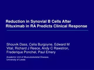
Reduction in Synovial B Cells After Rituximab in RA Predicts Clinical Response. Shouvik Dass, Celia Burgoyne, Edward M Vital, Richard J Reece, Andy C Rawstron, Frederique Ponchel, Paul Emery Academic Unit of Musculoskeletal Disease, University of Leeds. Selective B Cell Therapy in RA.

E N D
Reduction in Synovial B Cells AfterRituximab in RA Predicts Clinical Response Shouvik Dass, Celia Burgoyne, Edward M Vital, Richard J Reece, Andy C Rawstron, Frederique Ponchel, Paul Emery Academic Unit of Musculoskeletal Disease, University of Leeds
Selective B Cell Therapy in RA • Rituximab appears to deplete B cells effectively in peripheral blood – although variable depletion has been shown • Considerable variation exists in patients’ responses • B cell niches elsewhere – bone marrow, lymph node, synovium • Synovium – organised lymphoid infiltrates are seen in RA joints, with accumulation of plasma cells • Peripheral B cells eventually return but the relationship of this to disease activity & synovial status is unknown • We analysed synovial tissue before and 6 months after rituximab; compared changes in peripheral blood and synovium to clinical outcomes
Patients & Methods • Pilot study; initial results from first 10 patients • All RF+ve RA, inadequate responders (inefficacy & toxicity) or contraindications to anti-TNF therapy • All received RTX 1g x 2, Days 1 & 15 (preceded by methylprednisolone) • Clinical response assessed by EULAR response criteria every 3 months • Peripheral blood B-cell analysis by highly sensitive flow cytometry at baseline, Day 15 then 1,3,6 months1 - conventional limit of depletion: 0.005 x 10⁹/L - highly sensitive limit: 0.0001x 10⁹/L • Synovial biopsy with immunohistochemistry at baseline and 6 months 1Dass et al, ACR 2006,abstract no. 2121
Immunohistochemistry Method • Pre- and post-RTX synovial biopsies collected by knee arthroscopy, then frozen at -80°C • Cut 4m sections • Stained with primary antibodies – CD19, CD68, CD3 • 3 stage immunohistochemistry • Semi-quantitative scoring
Synovial CD19 Pre-rituximab Post-rituximab
Semi-quantitative Scoring • Sublining layer: B cells (or other rare cells) 0 = 0 – 10% 0 = 0% 1 = 10 – 25% 1 = 1 – 25% 2 = 25 – 50% 2 = 25 – 50% 3 = 50 – 75% 3 = 50 – 75% 4 = 75 – 100% 4 = 75 – 100% • Lining layer thickness: 1 = 1 – 2 cells 2 = 3 – 4 cells 3 = 5 – 6 cells 4 = 7+ cells Calculate mean score per field
Synovial B Cell Scores pre- and 6 Months Post-treatment with RTX 2.0 1.5 1.0 0.5 0.0 Patients exhibiting synovial B cell depletion
Synovial B Cell Scores Pre- and 6 Months Post-treatment with RTX 2.0 1.5 1.0 0.5 0.0 Patients exhibiting synovial B cell depletion Patients not exhibiting synovial B cell depletion
Greater Reduction in CD68 in Patients with Greater Reduction of CD19
Change in Ultrasound Synovitis U/S Grey Scale U/S Power Doppler
EULAR Response Rate is Better ifSynovial B Cells are Depleted *p 0.04 vs depleters Percent patients with good or moderate response * * months months Depleters Non-Depleters
High Sensitivity Flow Cytometry Reveals Incomplete Depletion and Earlier Return of B Cells After Therapy Patients with detectable B cells At 2 weeks, highly sensitive methods reveal large differences in B cell depletion. High sensitivity also identified return of B cells earlier than standard techniques 100 90 80 70 60 Patients (%) 50 40 30 20 10 0 Pre 2 weeks 4 weeks 3 months 6 months 9 months Standard High Sensitivity
B Cells – Peripheral & Synovial – and Clinical Response At 6 months: • 9/10 had detectable peripheral B cells • 7 of these patients still had synovial reduction
Level of Initial B Cell Depletion (Day 15) in PB Correlates with Later Synovial B Cell Expression
Summary • Varying levels of B cell depletion are seen in synovium: - the depth of depletion correlates with initial peripheral blood depletion • Varying clinical responses which correlate with synovial depletion at 6 months • Synovial repletion appears to be delayed compared to peripheral blood
Future Considerations & Questions • Early data with small numbers – few with poorer outcomes • What happens in synovium between 0 and 6 months? – patterns of depletion and repletion • What is the relationship between peripheral blood and synovial B cells and return of disease? • may be related to patterns of returning subsets in periphery • may be that after RTX, synovium is a less friendly environment for B cells • Effect of rituximab on other cell types and synovial milieu
Conclusions • B cell depletion after rituximab is variable • It differs between peripheral blood and synovium • Depth of initial peripheral depletion correlates with depth of later synovial depletion • Synovial reduction is maintained despite peripheral repletion • Greater synovial depletion at 6 months appears to result in better clinical responses
Acknowledgments • Research Nurses – Christine Thomas, Claire Brown • Technicians - Karen Henshaw & Diane Corscadden • Haematological Malignancy Diagnostic Service, Leeds