Download
1 / 24
240 likes | 254 Views
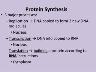
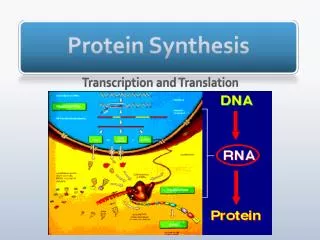
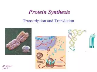
Protein synthesis. Genetic code. The genetic code is the way in which the nucleotide sequence in mRNA ( or DNA ) specifies the amino acid sequence in protein. Their nucleotide sequences are always written from the 5'-end to the 3'-end.

E N D
Genetic code • The genetic code is the way in which the nucleotide sequence in mRNA ( or DNA ) specifies the amino acid sequence in protein. • Their nucleotide sequences are always written from the 5'-end to the 3'-end. • The four nucleotide bases are used to produce the three-base codons. There are, therefore, 64 different combinations of bases, taken three at a time.
How to translate a codon • The genetic code table (or “dictionary”) can be used to translate any codon and, thus, to determine which amino acids are coded for by an mRNA sequence. For example, the codon 5'-AUG-3' codes for methionine • Sixty-one of the 64 codons code for the 20 common amino acids.
Termination (“stop” or “nonsense”) codons: • Three of the codons, UAG, UGA, and UAA, do not code for amino acids, but rather are termination codons. • When one of these codons appears in an mRNA sequence, synthesis of the polypeptide coded for by that mRNA stops.
Characteristics of the genetic code Specificity. Universal. Degeneracy. Nonoverlapping and commaless.
Consequences of altering the nucleotide sequence: • Changing a single nucleotide base on the mRNA chain (a “point mutation”) can lead to any one of three results: • Silent mutation: • 2. Missense mutation: • 3. Nonsense mutation:
A. Amino acids: All the amino acids that eventually appear in the finished protein must be present at the time of protein synthesis • B. Transfer RNA: At least one specific type of tRNA is required for each amino acid. • Amino acid attachment site: Each tRNA molecule has an attachment site for a specific (cognate) amino acid at its 3'-end. • Anticodon: Each tRNA molecule also contains a three-base nucleotide sequence—the anticodon—that pairs with a specific codon on the mRNA.
How many tRNA molecules are there? Wobble rules Unusual base
C. Aminoacyl-tRNA synthetases : This family of enzymes is required for attachment of amino acids to their corresponding tRNAs.. The extreme specificity of the synthetase in recognizing both the amino acid and its cognate tRNA contributes to the high fidelity of translation of the genetic message. In addition, the synthetases have a “proofreading” or “editing” activity that can remove amino acids from the enzyme or the tRNA molecule. D. Messenger RNA: The specific mRNA required as a template for the synthesis of the desired polypeptide chain must be present.
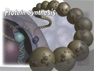
E. Functionally competent ribosomes : Ribosomes are large complexes of protein and ribosomal RNA They consist of two subunits one large and one. The small ribosomal subunit binds mRNA and is responsible for the accuracy of translation by ensuring correct base-pairing between the codon in the mRNA and the anticodon of the tRNA. The large ribosomal subunit catalyzes formation of the peptide bonds that link amino acid residues in a protein.
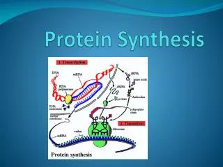
STEPS IN PROTEIN SYNTHESIS • The process of protein synthesis translates the three-letter alphabet of nucleotide sequences on mRNA into the 20-letter alphabet of amino acids that constitute proteins. • The mRNA is translated from its 5'-end to its 3'-end, producing a protein synthesized from its amino-terminal end to its carboxyl terminal end. • Prokaryotic mRNAs often have several coding regions, that is, they are polycistronic. • Each coding region has its own initiation and termination codon and produces a separate species of polypeptide. • In contrast, each eukaryotic mRNA has only one coding region, that is, it is monocistronic. • The process of translation is divided into three separate steps: initiation, elongation, and termination.
One important difference is that translation and transcription are coupled in prokaryotes, with translation starting before transcription is completed. • Coupling is a consequence of the lack of a nuclear membrane in prokaryotes.
Shine-Dalgarno sequence Complementary binding between prokaryotic mRNA Shine-Dalgarno sequence and 16S rRNA. • In E. coli, a purine-rich sequence of nucleotide bases, is located six to ten bases upstream of the initiating AUG codon on the mRNA molecule—that is, near its 5'-end. • Eukaryotic messages do not have SD sequences. • In eukaryotes, the 40S ribosomal subunit binds close to the cap structure at the 5-end of the mRNA and moves down the mRNA until it encounters the initiator AUG.
B- Elongation • Elongation of the polypeptide chain involves the addition of amino acids to the carboxyl end of the growing chain. • Delivery of the aminoacyl-tRNA whose codon appears next on the mRNA template in the ribosomal A site is facilitated in E. coli by elongation factors EF-Tu-GTP and EF-Ts, and requires GTP hydrolysis. • After the peptide bond has been formed, what was attached to the tRNA at the P site is now linked to the amino acid on the tRNA at the A site. • The ribosome then advances three nucleotides toward the 3'-end of the mRNA. This process is known as translocation. • Translocation causes movement of the uncharged tRNA from the P to the E site, and movement of the peptidyl -tRNA from the A to the P site. • The process is repeated until a termination codon is encountered.
C- Termination • Termination occurs when one of the three termination codons moves into the A site. These codons are recognized in E. coli by release factors: RF-1, which recognizes the termination codons UAA and UAG, and RF-2, which recognizes UGA and UAA. • The binding of these release factors results in hydrolysis of the bond linking the peptide to the tRNA at the P site, causing the nascent protein to be released from the ribosome. • A third release factor, RF-3-GTP then causes the release of RF-1 or RF-2 as GTP is hydrolyzed .
An overview of the main events in translation. Translation is initiated by the pairing of an mRNA and tRNA on the ribosome. In elongation, the ribosome moves along the mRNA, matching tRNAs to each codon and catalyzing peptide bond formation. Translation terminates at a stop codon, and the ribosomal subunits are released for another round of synthesis
CO- and Posttranslational modification of polypeptide chains • Many polypeptide chains are covalently modified, either while they are still attached to the ribosome (cotranslational) or after their synthesis has been completed (posttranslational). • These modifications may include removal of part of the translated sequence, or the covalent addition of one or more chemical groups required for protein activity.
Some types of posttranslational modifications are listed below. A. Trimming • Many proteins destined for secretion from the cell are initially made as large, precursor molecules that are not functionally active. • Portions of the protein chain must be removed by specialized endoproteases, resulting in the release of an active molecule. • The cellular site of the cleavage reaction depends on the protein to be modified. B. Covalent attachments • Proteins may be activated or inactivated by the covalent attachment of a variety of chemical groups. • Phosphorylation • Glycosylation • Hydroxylation
C. Protein folding • Proteins must fold to assume their functional state. Folding can be spontaneous (as a result of the primary structure), or facilitated by proteins known as “chaperones” . • D. Protein degradation • Proteins that are defective, for example, misfolded, or destined for rapid turnover are often marked for destruction by ubiquitination— the attachment of chains of a small, highly conserved protein, called ubiquitin. • Proteins marked in this way are rapidly degraded by a cellular component known as the proteasome, which is a macromolecular, ATP-dependent, proteolytic system located in the cytosol.