Campylobacter
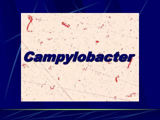
Campylobacter . Campylobacter. Among the most widespread cause of infection in the world. Cause both diarrheal and systemic diseases Campylobacter jejuni. Typical Organisms. Gram-negative rods with comma, S, or “ gull-wing ” shapes. Motive, with a single polar flagellum

Campylobacter
E N D
Presentation Transcript
Campylobacter • Among the most widespread cause of infection in the world. • Cause both diarrheal and systemic diseases • Campylobacter jejuni
Typical Organisms • Gram-negative rods with comma, S, or “gull-wing” shapes. • Motive, with a single polar flagellum • No spore & no capsule
Culture • An atmosphere with reduced O2 (5% O2) with added CO2 (10% CO2) • At 42 ℃ (for selection) • Several selective media can be used (eg, Skirrow’s medium) • Two types of colonies: watery and spreading round and convex
Virulence Factor • Lipopolysaccharides (LPS) with endotoxic activity • Cytopathic extracellular toxins and enterotoxins have been found
Pathogenesis • The infection by oral route from food, drink, or contact with infected animals or animal products(Milk,meat products ). • Susceptible to gastric acid (about 104 organisums) • Multiply in the small intestine invade the epithium produce inflammation cause bloody stools • Occasionally, the bloodstream is invaded
Campylobacter - symptoms • Incubation: 4-8d • Acute enteritis: 1w, stools remain positive for 3 w • Acute colitis • Acute abdominal pain • Bacteremia: <1% C. jejuni • Septic abortion • Reactive arthritis • diarrhea • malaise • fever • abdominal pain • usually self-limiting • antibiotics occassionally • bacteremia • small minority
Diagnostic Laboratory Tests • Specimens: Diarrheal stools • Smears: Gram-stained smears of stool may show the typical “gull-shaped” rods. • Culture: (have been described above)
Control • The source of infection may be food (eg, milk, under-cooked fowl) or contract with infected animals or humans and their excreta.
Helicobacter pylori Curved bacilli – Former name - Campylobacter pylori, H. pylori
Helicobacter pylori • Helicobacter pylori is the prototype organism in this group. It is associated with antral gastritis, gastric ulcers, and gastric carcinoma.
Microbiology Gram negative rod, curved, Very Motile corkscrew motion Microaerophilic, use amino acids and fatty acids rather than carbohydrates to obtain energyneeds 10% CO2 and 5% O2 Urease production Catalase production Oxidase positive Growth at 370C, not 250C or 420C
Virulence factors • vacA (vacuolationg associated) cytotoxin, Pathogenicity island: cag, cytotoxin associated gene A+genes related to bacterial secretion • Cag+ HP is much more associated with peptic ulcer disease than Cag(--) HP.
Pathogenesis Motility – it moves into the mucus and produces adhesins on gastric epithelial cells (not intestinal epithelial cells) Urease production, breaks down the urea to ammonia which buffers the pH around the bacterium. Persists, escape defense mechanisms – SOD, catalase, Urease. Breack down free radicals
Pathogenesis • H pylori invade the epithelial cell surface to a certain degree • Toxins and LPS may damage the mucosal cells • NH3 produced by the urease activity may also damage the cells
Epidemiology • Prevalence related to socioeconomic level during childhood. • Infection occurs in childhood, persists for decades • Prevalence among adults – 20%-100% • Source – stomach of humans • Mode of transmission? Fecal-oral? Oral-oral? Vomiting and aerosols ? • Incidence of HP colonization is declining in developed countries
Epidemiology • Under age 30 <20% At age 60 40-60% • In developing countries >80% in adults • Acute epidemics of gastritis suggest a common source for H pylori.
Clinical features • Acute acquisition - nausea, vomiting, abdominal pain • last for 1w, later –gastritis. • Persistent colonization - after acquisition, persist for years. Asymptomatic. • Duodenal ulcer • more than 90% with DU - carry HP. • antimicrobial therapy response, eradication of HP - less recurrences
Gastric ulcer - 50-80% HP • Gastric carcinoma -HP induces gastritis, gastritis is risk factor for Carcinoma. • Gastric lymphoma - MALToma: mucosa associated lymphoid tumors, strong association with HP. Stage 1 is cured by antibiotics. • Esophageal diseases - HP protects against: gastroesophageal reflux, Barrette's esophagus and carcinoma of esophagus.
Immunity • An IgM antibody response to he infection is developed • Subsequently, IgG and IgA are produced
Laboratory diagnosis Endoscopy and biopsy. Urease detection Culture Urea breath test - samples of breath air are collected by having the patient blow into a tube before and 30 min after ingestion of 13C-labeled urea, rapid, noninvasive, for assessing response 4-8w post therapy, expensive but non invasive!! Serology
Principles of therapy • Combination chemotherapy • Some drugs are effective in vitro, not in vivo - due to acidic pH - erythromycin • Resistance - not to bismuth salts or tetracyclines, 10-30% to metronidazole, • Response - 1 month after cessation of therapy for breath test or biopsy, 6 month for serology
Principles of therapy • Triple therapy: Bismuth+metronidazole+amoxicillin: eradication 60-90%, tetracyclines, macrolides - clarithromycin • PPI proton pump inhibitors therapy: omeprazolone lansoprazole: inhibit HP, urease, acid • PPI+amoxicillin+clarithromycin or metronidazole • PPI+ Bismuth+metronidazole+amoxicillin-very effective
PSEUDOMONAS 假单孢菌属
Common Characteristics • Gram-negative • Motile • Aerobic rod • Some produce water-soluble pigments • Widely in soil, water, plants and animals • More than 200 (up to now)
Pseudomonas aeruginosa • Widely distributed in nature • Frequently present in small numbers in the normal intestinal flora and on the skin • Commonly present in moist environments in hospitals • It is primarily a nosocomial pathogen
Typical Organisms • Gram-negative rod ---- 0.6×2 μm • Unipolar flagellum (1~3) ---- actively mobile • Occurs as single bacteria, in pairs, and occasionally in short chain • Capsule • Pili in strains obtained from clinical specimens
Culture • Grow readily on many types of culture media • Smooth and round colonies • Multiple colony types in one culture • Fluorescent greenish color • Sometimes produce a sweet or grape-like or corn taco-like odor
Culture • Obligate aerobic • Grow well at 37~42℃and no growth at 4℃ • Produce water-soluble pigments Pyocyanin; Pyoverdin; Pyorubin; Pyomelanin • Produce hemolysin • Oxidase-positive • Ferment glucose but not other carbohydrates
Virulence Determinants • Adhesins fimbriae (N-methyl-phenylalanine pili) polysaccharide capsule (glycocalyx) alginate slime (biofilm) • Invasins elastase alkaline protease hemolysins (phospholipase and lecithinase) cytotoxin (leukocidin) siderophores and siderophore uptake systems pyocyanin diffusible pigment
Virulence Determinants • Motility/chemotaxis Flagella • Toxins Exoenzyme S Exotoxin A Lipopolysaccharide • Antiphagocytic surface properties Capsules, slime layers LPS • Defense against serum bactericidal reaction Slime layers,capsules LPS Protease enzymes
Virulence Determinants • Defense against immune responses Capsules, slime layers Protease enzymes • Genetic attributes Genetic exchange by transduction and conjugation Inherent (natural) drug resistance R factors and drug resistance plasmids • Ecologic criteria Adaptability to minimal nutritional requirements Metabolic diversity Widespread occurrence in a variety of habitats
Inhibition of protein synthesis in susceptible cells ----Toxin A • The resultant ADP-ribosyl-EF-2 complex is inactive in protein synthesis. • This intracellular mechanism of action of toxin A is identical to that of diphtheria toxin fragment A .
Disease caused by Pseudomonas aeruginosa • Endocarditis • Respiratory infections • Bacteremia • Central Nervous System infections • Ear infections including external otitis • Eye infections • Bone and joint infections • Urinary tract infections • Gastrointestinal infections • Skin and soft tissue infections, including wound infections, pyoderma and dermatitis
Who are at risk? • People with cystic fibrosis • Burn victims • Individuals with cancer • Patients requiring extensive stays in intensive care units
Diagnosis • Isolation and laboratory identification. blood agar plates eosin-methylthionine blue agar. • Gram morphology, • Inability to ferment lactose • Positive oxidase reaction • Fruity odor • Ability to grow at 4 2 ℃ • Fluorescence under ultraviolet radiation helps in early identification of P aeruginosa colonies and also is useful in suggesting its presence in wounds.
Control and Treatment • The spread of Pseudomonas is best controlled by cleaning and disinfecting medical equipment. • In burn patients, topical therapy of the burn with antimicrobial agents such as silver sulfadiazine, coupled with surgical debridement, has markedly reduced sepsis. • Susceptibility testing is essential. • The combination of gentamicin and carbenicillin can be very effective in patients with acute P aeruginosa infections.
Review • General characteristics: Gram negative rod, unipolar flagellum, actively motile; produce diffusible pigments -- pyocyanin,gluorescin and pyorubin; aerobic, produce hemolysin. • Pathogenicity: cause suppurative infections in burn, trauma, etc. Endotoxin: main pathogenic substance Exotoxin A Extracellular enzymes:phospholipase, proteinase, etc. • Bacteriological diagnosis: Specimens Culture and identification Unusual bacteria
Common Characteristics • Small, gram-negative • Pleomorphic • Require enrich media (usually containing blood for isolation) • No flagellum, no spore • Divided into 17 species according to different requirement to X and V factor
Haemophilus • Small Gram-negative coccobacilli, facultative anaerobes, non motile • often resemble cocci, eg pneumococci, • most non-encapsulated strains --- virulent forms encapsulated • fastidious (require blood factors)X factor = hematinV factor = NAD • Organisms: H. influenzae: H. ducreyi --( soft chancre); H. aegypticus -- (purulent conjunctivitis)
Characteristics and growth requirements of some haemophilus species • X=heme; V=nicotinamide-adenine dinucleotide