Blood Composition and Function
Blood Composition and Function. General Composition of Blood Plasma Formed Elements Erythrocytes Leukocytes Hematopoesis Hemostasis (Clotting) Transfusion and Blood Typing Developmental Aspects of Blood. Blood Composition. The only fluid tissue in the human body

Blood Composition and Function
E N D
Presentation Transcript
Blood Composition and Function • General Composition of Blood • Plasma • Formed Elements • Erythrocytes • Leukocytes • Hematopoesis • Hemostasis (Clotting) • Transfusion and Blood Typing • Developmental Aspects of Blood
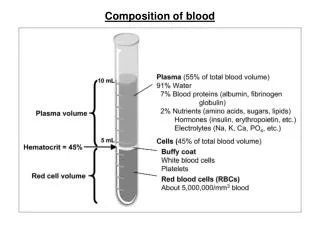
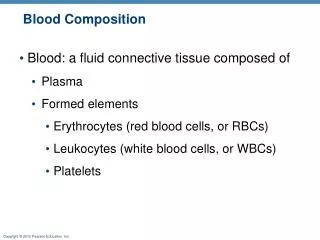
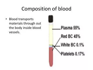
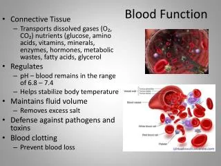
Blood Composition • The only fluid tissue in the human body • Classified as a connective tissue • Living cells = formed elements • Non-living matrix = plasma • Color range • Oxygen-rich blood is scarlet red • Oxygen-poor blood is dull red • pH must remain between 7.35–7.45 • Blood temperature is slightly higher than body temperature Hematocrit: Percent of blood volume that is RBCs • 47% ± 5% for males • 42% ± 5% for females Figure 10.1
Blood Composition and Function • General Composition of Blood • Plasma • Formed Elements • Erythrocytes • Leukocytes • Hematopoesis • Hemostasis (Clotting) • Transfusion and Blood Typing • Developmental Aspects of Blood
Erythrocytes (Red Blood Cells) • Main function is to carry oxygen • Biconcave disks • Essentially bags of hemoglobin; few organelles • Anucleate (no nucleus) • Outnumber white blood cells 1000:1 • Contain the plasma membrane protein spectrin and other proteins • Major factor contributing to blood viscosity
Hemoglobin • Molecular structure • Protein globin: two alpha and two beta chains • Heme pigment bonded to each globin chain • Iron atom in each heme can bind reversibly to one O2 molecule • Each Hb molecule can transport four O2
Sickle Cell Hemoglobin Causes Clotting • Normal hemoglobin = HbA • Sickle cell hemoglobin = HbS (mutation in single amino acid - missense)
Pleiotropic Effects Seen if HbS present HbAHbA = normal HbAHbS = sickle cell trait HbSHbS = sickle cell disease/anemia
Fetal Hemoglobin Binds O2 More Avidly • Fetal circulation must extract oxygen from maternal circulation • Fetal hemoglobin (HbF) binds O2 more avidly than normal hemoglobin (HbA) does • Replaced by “adult” hemoglobin just after birth
Blood Composition and Function • General Composition of Blood • Plasma • Formed Elements • Erythrocytes • Leukocytes • Hematopoesis • Hemostasis (Clotting) • Transfusion and Blood Typing • Developmental Aspects of Blood
Leukocytes (White Blood Cells) Differential WBC count (All total 4800– 10,800/l) Formed elements Platelets Granulocytes Neutrophils (50–70%) Leukocytes Eosinophils (2–4%) Basophils (0.5–1%) Erythrocytes Agranulocytes Lymphocytes (25 – 45%) Monocytes (3 – 8%) Figure 17.9
Leukocyte Levels in the Blood • Normal levels are between 4,000 - 11,000 cells per millimeter • Abnormal leukocyte levels • Leukocytosis • Above 11,000 leukocytes/ml • Generally indicates an infection (bacterial, fungal, parasitic) • Also caused by some cancers, (including lymphocyte leukemia), hemhorrage, pneumonia • Leukopenia • Abnormally low leukocyte level (< 3,500/ml) • Commonly caused by certain drugs, radiation • Neutropenia (low neutrophil count) common in cancer patients receiving chemotherapy • HIV-AIDS, TB, malaria, influenza also cause leukopenia • Requires careful sanitation and “gowning up” for medical staff
Types of Leukocytes • Granulocytes • Granules in their cytoplasm can be stained • Include neutrophils, eosinophils, and basophils • Agranulocytes • Lack visible cytoplasmic granules • Include lymphocytes and monocytes Figure 10.4
Blood Composition and Function • General Composition of Blood • Plasma • Formed Elements • Erythrocytes • Leukocytes • Hematopoesis • Hemostasis (Clotting) • Transfusion and Blood Typing • Developmental Aspects of Blood
Hematopoiesis • Subdivided into erythropoesis and leukopoesis • Erythropoesis ccurs in red bone marrow • Erythropoesis Steps • Ribosome synthesis • Hemoglobin accumulation • Ejection of the nucleus and formation of reticulocytes • Reticulocytes then become mature erythrocytes Stem cell Committed cell Developmental pathway Phase 1 Ribosome synthesis Phase 2 Hemoglobin accumulation Phase 3 Ejection of nucleus Figure 17.5 Reticulo- cyte Erythro- cyte Proerythro- blast Early erythroblast Late erythroblast Normoblast Hemocytoblast
Fate of Erythrocytes • Unable to divide, grow, or synthesize proteins • Wear out in 100 to 120 days • When worn out, are eliminated by phagocytes in the spleen or liver • Iron is salvaged for reuse • Heme is degraded to yellow the pigment bilirubin • Liver secretes bilirubin (in bile)) into the intestines • Lost cells are replaced by division of hemocytoblasts
Control of Erythrocyte Production • Rate is controlled by a hormone (erythropoietin) • Kidneys produce most erythropoietin as a response to reduced oxygen levels in the blood (hypoxia) • Hypoxia can develop from hemorrhage, low O2 in air, insufficient hemoglobin • Homeostasis is maintained by negative feedback from blood oxygen levels
Platelets Important in Clotting • Small fragments of megakaryocytes • Formation is regulated by thrombopoietin • Blue-staining outer region, purple granules • Form a temporary platelet plug that helps seal breaks in blood vessels Stem cell Developmental pathway Hemocyto- blast Megakaryocyte Platelets
Blood Composition and Function • General Composition of Blood • Plasma • Formed Elements • Erythrocytes • Leukocytes • Hematopoesis • Hemostasis (Clotting) • Transfusion and Blood Typing • Developmental Aspects of Blood
Hemastasis: Blood Clotting 1 Step Vascular spasm • Smooth muscle contracts, causing vasoconstriction. 2 Step Platelet plug formation •Injury to lining of vessel exposes collagen fibers; platelets adhere. Collagen fibers •Platelets release chemicals that make nearby platelets sticky; platelet plug forms. Platelets 3 Step Coagulation •Fibrin forms a mesh that traps red blood cells and platelets, forming the clot. Fibrin Figure 17.13
Undesirable Clotting • Thrombus • A clot in an unbroken blood vessel • Can be deadly in areas like the heart • Embolus • A thrombus that breaks away and floats freely in the bloodstream • Can later clog vessels in critical areas such as the brain, causing a stroke (CVA)
Bleeding Disorders • Thrombocytopenia • Platelet deficiency • Even normal movements can cause bleeding from small blood vessels that require platelets for clotting • Hemophilia • Hereditary bleeding disorder • Normal clotting factors are missing or deficiency in Vit. K. mild hemophilia
Blood Composition and Function • General Composition of Blood • Plasma • Formed Elements • Erythrocytes • Leukocytes • Hematopoesis • Hemostasis (Clotting) • Transfusion and Blood Typing • Developmental Aspects of Blood
Blood Groups and Transfusions • Large losses of blood have serious consequences • Loss of 15 to 30 percent causes weakness • Loss of over 30 percent causes shock, which can be fatal • Transfusions are the only way to replace blood quickly • Transfused blood must be of the same blood group
Human Blood Groups • Blood contains genetically determined proteins • A foreign protein (antigen) on a donated RBC may be attacked by the immune system • Blood is “typed” by using antibodies that will cause blood with certain proteins to clump (agglutination)
Human Blood Groups • There are over 30 common red blood cell antigens • The most vigorous transfusion reactions are caused by ABO and Rh blood group antigens ABO Blood Groups and Alleles Components of Blood of this Type anti-A anti-B anti-B anti-A
Rh Blood Groups and Hemolytic Deaseas of Newborns • Named because of the presence or absence of one of eight Rh antigens (agglutinogen D) • Most Americans are Rh+ • The danger is only when the mother is Rh– and the father is Rh+,and the child inherits the Rh+ factor Dd dd This baby, if from the second or subsequent pregnancy, could have complications in an Rh- mother • Problems can occur in second and subsequent pregnancies when the baby’s Rh+ blood enters Rh– maternal circulation: erythroblastosis fetalis (hemolytic disease of the newborn) • RhoGAM injections contain anti-Rh antibodies that quickly clear the maternal circulation of Rh protein Dd dd
Blood Composition and Function • General Composition of Blood • Plasma • Formed Elements • Erythrocytes • Leukocytes • Hematopoesis • Hemostasis (Clotting) • Transfusion and Blood Typing • Developmental Aspects of Blood