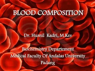
BLOOD COMPOSITION
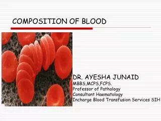
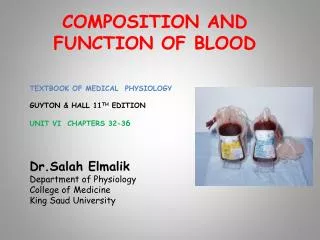
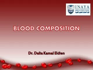
BLOOD COMPOSITION. Dr. Dalia Kamal Eldien. INTRODUCTION:.

BLOOD COMPOSITION
E N D
Presentation Transcript
BLOOD COMPOSITION Dr. Dalia KamalEldien
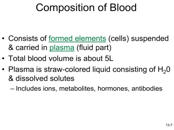
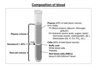
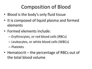
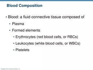
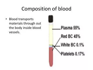
INTRODUCTION: • Blood is composed of fluid plasma and cells. There are different kinds of cells (occasionally called corpuscles); which constitute about 45% of whole blood. The other 55% is blood plasma, a fluid that is the blood's liquid medium, appearing yellow in color.
The normal pH of human arterial blood is approximately 7.40 (normal range is 7.35-7.45), a weak alkaline solution. Blood is about 7% of the human body weight, so the average adult has a blood volume of about 5 liters, of which 2.7-3 liters is plasma.
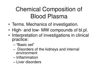
BLOOD PLASMA: • When the formed elements are removed from blood, a straw-colored liquid called plasma is left. Plasma is about 91.5% water and 8.5% solutes, most of which by weight (7%) are proteins. • Some of the proteins in plasma are also found elsewhere in the body, but those confined to blood are called plasma proteins.
Most plasma proteins are synthesized by the liver, including the albumins (54% of plasma proteins), globulins (38%), and fibrinogen (7%). • Other solutes in plasma include waste products, such as urea, uric acid, creatinine, ammonia, and bilirubin; nutrients; vitamins; regulatory substances such as enzymes and hormones; gasses; and electrolytes.
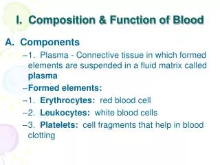
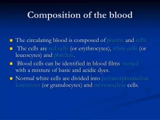
FORMED ELEMENTS: • The formed elements of the blood are broadly classified as red blood cells (erythrocytes), white blood cells (leucocytes) and platelets (thrombocytes).
RED BLOOD CELL • They are the most numerous cells in the blood. In adults, they are formed in the marrow of the bones. Mature red cells are non- nucleated and are shaped like flattened, bilaterally spheres, referred to as “biconcave disc” with a diameter 7.0-8.0 μm and thickness of 1.7-2.4 μm. In stained smears, only the flattened surfaces are observed.
They are primarily involved in tissue respiration. The red cells contain the pigment hemoglobin which has the ability to combine reversibly with 02. In the lungs, the hemoglobin in the red cell combines with 02 and releases it to the tissues of the body (where oxygen tension is low) during its circulation. Carbondioxide, a waste product of metabolism, is then absorbed from the tissues by the red cells and is transported to the lungs to be exhaled.
The red cell normally survives in the blood stream for approximately 120 days after which time it is removed by the phagocytic cells of the reticuloendothelial system. • Male: 4.7 to 6.1 million cells per microliter (cells/mcL) • Female: 4.2 to 5.4 million cells/mcL • Children: 4.6 to 4.8 million/uL
White Blood Cells • They are a heterogeneous group of nucleated cells that are responsible for the body’s defenses and are transported by the blood to various tissues where they exert their physiologic role, e.g. phagocytosis. WBCs are present in normal blood in smaller number than the red blood cells (5.0-10.0 x 10³ /μl in adults). Their production is in the bone marrow and lymphoid tissues (lymph nodes, lymph nodules and spleen).
There are five types of WBC each with a characteristic morphologic appearance and specific physiologic role. These are: • Polymorphonuclear leucocytes/granulocytes: Neutrophils, Eosinophils & Basophiles. • Mononuclear leucocytes: Lymphocytes & Monocytes.
Polymorphonuclear Leucocytes • Neutrophils: Their size ranges from 10-12μm in diameter. They are capable of amoeboid movement. There are 2-5 lobes to their nucleus that stain purple violet. The cytoplasm stains light pink with pinkish dust like granules. Normal range: 2.0-7.5 x 10³/μl. Their number increases in acute bacterial infections.
Eosinophils: have the same size as neutrophils or may be a bit larger (12-14 μm).There are two lobes to their nucleus in a "spectacle" arrangement. Their nucleus stains a little paler than that of neutrophils. The cytoplasm contains many, large, round/oval orange pink granules. They are involved in allergic reactions and in combating helminthic infections. Normal range: 40-400/μl. Increase in their number (eosinophilia) is associated with allergic reactions and helminthiasis.
Basophils: Their size ranges from 10-12 μm in diameter. Basophils have a kidney shaped nucleus frequently obscured by a mass of large deep purple/blue staining granules. Their cytoplasmic granules contain heparin and histamine that are released at the site of inflammation. Normal range: 20-200/μl. Basophilia is rare except in cases of chronic myeloid leukemia.
Mononuclear leucocytes • Lymphocytes: there are two varieties: • Small Lymphocytes: Their size ranges from 7-10μm in diameter. Small lymphocytes have round, deep-purple staining nucleus which occupies most of the cell. There is only a rim of pale blue staining cytoplasm. They are the predominant forms found in the blood. • Large Lymphocytes: Their size ranges from 12-14μm in diameter. They have a little paler nucleus than small lymphocytes. They have more plentiful cytoplasm that stains pale blue and may contain a few reddish granules. The average number of lymphocytes in the peripheral blood is 2500/μl. Lymphocytosis is seen in viral infections especially in children.
Monocytes: are the largest white cells measuring 14-18μm in diameter. They have a centrally placed, large and (horseshoe) shaped nucleus that stains pale violet. Their cytoplasm stains pale grayish blue and contains reddish blue dust-like granules and a few clear vacuoles. They are capable of ingesting bacteria. Normal range: 700-1500/μl. Monocytosis is seen in bacterial infections. (e.g. tuberculosis) and protozoan infections.
Platelets • These are small, non nucleated, round/oval cells that stain pale blue and contain many pink granules. Their size ranges 1-4 µm in diameter. They are produced in the bone marrow by fragmentation of cells called megakaryocytes which are large and multinucleated cells. Their primary function is preventing blood loss from hemorrhage.
When blood vessels are injured, platelets rapidly adhere to the damaged vessel and with one another to form a platelet plug. During this process, the soluble blood coagulation factors are activated to produce a mesh of insoluble fibrin around the clumped platelets. This assists and strengthens the platelet plug and produces a blood clot which prevents further blood loss. Normal range: 150-400 x 103/µl.
FUNCTION OF THE BLOOD • Transportation: transport O2 form lungs to body’s cells and CO2 from the cells to the lungs. It also carries nutrients from the GIT to the cells, heat and waste products away from cells and hormones form endocrine glands to other body cells. • Regulation: regulates pH through buffers. It also adjusts body temperature through the heat-absorbing and coolant properties of its water content and its variable rate of flow through the skin. Blood osmotic pressure also influences the water content of cells.
Protection: The clotting mechanism protects against blood loss, and certain phagocytic white blood cells or specialized plasma proteins such as antibodies & interferon protect against foreign microbes and toxins.