Download

1 / 7
70 likes | 198 Views
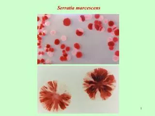
A study was performed to evaluate the impact of biofield treatment on phenotyping and genotyping characteristics of S. marcescens. Visit here for more details.<br>

E N D
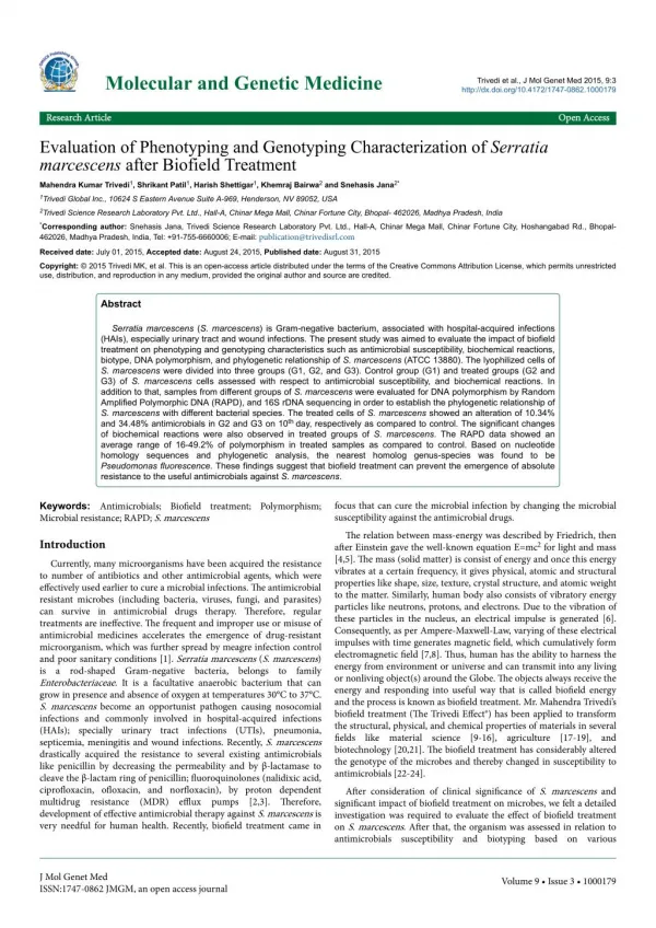
Molecular and Genetic Medicine Trivedi et al., J Mol Genet Med 2015, 9:3 http://dx.doi.org/10.4172/1747-0862.1000179 Research Article Research Article Open Access Open Access Evaluation of Phenotyping and Genotyping Characterization of Serratia marcescens after Biofield Treatment Mahendra Kumar Trivedi1, Shrikant Patil1, Harish Shettigar1, Khemraj Bairwa2 and Snehasis Jana2* 1Trivedi Global Inc., 10624 S Eastern Avenue Suite A-969, Henderson, NV 89052, USA 2Trivedi Science Research Laboratory Pvt. Ltd., Hall-A, Chinar Mega Mall, Chinar Fortune City, Bhopal- 462026, Madhya Pradesh, India *Corresponding author: Snehasis Jana, Trivedi Science Research Laboratory Pvt. Ltd., Hall-A, Chinar Mega Mall, Chinar Fortune City, Hoshangabad Rd., Bhopal- 462026, Madhya Pradesh, India, Tel: +91-755-6660006; E-mail: publication@trivedisrl.com Received date: July 01, 2015, Accepted date: August 24, 2015, Published date: August 31, 2015 Copyright: © 2015 Trivedi MK, et al. This is an open-access article distributed under the terms of the Creative Commons Attribution License, which permits unrestricted use, distribution, and reproduction in any medium, provided the original author and source are credited. Abstract Serratia marcescens (S. marcescens) is Gram-negative bacterium, associated with hospital-acquired infections (HAIs), especially urinary tract and wound infections. The present study was aimed to evaluate the impact of biofield treatment on phenotyping and genotyping characteristics such as antimicrobial susceptibility, biochemical reactions, biotype, DNA polymorphism, and phylogenetic relationship of S. marcescens (ATCC 13880). The lyophilized cells of S. marcescens were divided into three groups (G1, G2, and G3). Control group (G1) and treated groups (G2 and G3) of S. marcescens cells assessed with respect to antimicrobial susceptibility, and biochemical reactions. In addition to that, samples from different groups of S. marcescens were evaluated for DNA polymorphism by Random Amplified Polymorphic DNA (RAPD), and 16S rDNA sequencing in order to establish the phylogenetic relationship of S. marcescens with different bacterial species. The treated cells of S.marcescens showed an alteration of 10.34% and 34.48% antimicrobials in G2 and G3 on 10th day, respectively as compared to control. The significant changes of biochemical reactions were also observed in treated groups of S. marcescens. The RAPD data showed an average range of 16-49.2% of polymorphism in treated samples as compared to control. Based on nucleotide homology sequences and phylogenetic analysis, the nearest homolog genus-species was found to be Pseudomonas fluorescence. These findings suggest that biofield treatment can prevent the emergence of absolute resistance to the useful antimicrobials against S. marcescens. Keywords: Antimicrobials; Biofield treatment; Polymorphism; Microbial resistance; RAPD; S. marcescens focus that can cure the microbial infection by changing the microbial susceptibility against the antimicrobial drugs. The relation between mass-energy was described by Friedrich, then after Einstein gave the well-known equation E=mc2 for light and mass [4,5]. The mass (solid matter) is consist of energy and once this energy vibrates at a certain frequency, it gives physical, atomic and structural properties like shape, size, texture, crystal structure, and atomic weight to the matter. Similarly, human body also consists of vibratory energy particles like neutrons, protons, and electrons. Due to the vibration of these particles in the nucleus, an electrical impulse is generated [6]. Consequently, as per Ampere-Maxwell-Law, varying of these electrical impulses with time generates magnetic field, which cumulatively form electromagnetic field [7,8]. Thus, human has the ability to harness the energy from environment or universe and can transmit into any living or nonliving object(s) around the Globe. The objects always receive the energy and responding into useful way that is called biofield energy and the process is known as biofield treatment. Mr. Mahendra Trivedi’s biofield treatment (The Trivedi Effect®) has been applied to transform the structural, physical, and chemical properties of materials in several fields like material science [9-16], agriculture [17-19], and biotechnology [20,21]. Thebiofield treatment has considerably altered the genotype of the microbes and thereby changed in susceptibility to antimicrobials [22-24]. Introduction Currently, many microorganisms have been acquired the resistance to number of antibiotics and other antimicrobial agents, which were effectively used earlier to cure a microbial infections. The antimicrobial resistant microbes (including bacteria, viruses, fungi, and parasites) can survive in antimicrobial drugs therapy. Therefore, regular treatments are ineffective.The frequent and improper use or misuse of antimicrobial medicines accelerates the emergence of drug-resistant microorganism, which was further spread by meagre infection control and poor sanitary conditions [1]. Serratia marcescens (S. marcescens) is a rod-shaped Gram-negative bacteria, belongs to family Enterobacteriaceae. It is a facultative anaerobic bacterium that can grow in presence and absence of oxygen at temperatures 30°C to 37°C. S. marcescens become an opportunist pathogen causing nosocomial infections and commonly involved in hospital-acquired infections (HAIs); specially urinary tract infections (UTIs), pneumonia, septicemia, meningitis and wound infections. Recently, S. marcescens drastically acquired the resistance to several existing antimicrobials like penicillin by decreasing the permeability and by β-lactamase to cleave the β-lactam ring of penicillin; fluoroquinolones (nalidixic acid, ciprofloxacin,ofloxacin, and norfloxacin), by proton dependent multidrug resistance (MDR) efflux pumps [2,3]. Therefore, development of effective antimicrobial therapy against S. marcescens is very needful for human health. Recently, biofield treatment came in After consideration of clinical significance of S. marcescens and significant impact of biofield treatment on microbes, we felt a detailed investigation was required to evaluate the effect of biofield treatment on S. marcescens. After that, the organism was assessed in relation to antimicrobials susceptibility and biotyping based on various J Mol Genet Med ISSN:1747-0862 JMGM, an open access journal Volume 9 • Issue 3 • 1000179
Citation: Trivedi MK, Patil S, Shettigar H, Bairwa K, Jana S (2015) Evaluation of Phenotyping and Genotyping Characterization of Serratia marcescens after Biofield Treatment. J Mol Genet Med 9: 179. doi:10.4172/1747-0862.1000179 Page 2 of 7 biochemical reactions. We also explored the genotyping of this organism using polymerase chain reaction (PCR) based methodologies of randomly amplified polymorphic DNA (RAPD) and 16S rDNA sequencing techniques. To the best of our knowledge, this is the first report that explores the impact of biofield treatment on S. marcescens. glucose, hydrogen sulfide, indole, inositol, kanamycin, lysine, malonate, melibiose, nitrate, oxidation, galactosidase, ornithine, oxidase, raffinose, rhamnose, sorbitol, sucrose, tartrate, tobramycin, urea, and Voges-Proskauer. Biotype number Materials and Methods The biotype number of S. marcescens was determined by MicroScan Walk-Away® processed panel data utilizing biochemical reactions data [25]. Two vials of S. marcescens [American Type Culture Collection (ATCC) 13880] were procured from MicroBioLogics, Inc., USA, in sealed packs, and stored as per the recommended storage conditions until further use. The anti-microbial susceptibility, biochemical reactions, and biotype number were evaluated on MicroScan Walk- Away® (Dade Behring Inc., West Sacramento, CA) using Negative Breakpoint Combo 30 (NBPC30). DNA Fingerprinting by RAPD analysis (using Ultrapure Genomic DNA Prep Kit; Cat KT 83) and the 16S rDNA sequencing studies were carried out using Ultrapure Genomic DNA Prep Kit; Cat KT 83 (Bangalore Genei, India). All the tested antimicrobials, biochemicals and other reagents were procured from Sigma-Aldrich. Random Amplified Polymorphic DNA (RAPD) analysis Three inoculums (one for control and other two for treatment named as treatment A and B) were prepared of S. marcescens samples. Two inoculums (treatment samples A and B) were subjected to Mr. Trivedi's biofield treatment. After that, the treated samples were sub- cultured by taking 1% inoculum and inoculated to fresh 5 mL medium and labeled as treatment A-1 and treatment B-1, respectively. All samples were incubated at 37°C with 160 rpm for 18 h. Subsequently, the cultures were spun down, and genomic DNA was isolated for control and treated samples using Genomic DNA Prep Kit (Bangalore Genei, India). RAPD was performed with all samples of S. marcescens using five RAPD primers, which were labelled as RBA8A, RBA13A, RBA20A, RBA10A and RBA15A. The PCR mixture contained 2.5 µL each of buffer, 4.0 mM each of dNTP, 2.5 μM each of primer, 5.0 μL each of genomic DNA, 2 U each of Taq polymerase, 1.5 μL of MgCl2 and 9.5 μL of water in a total of 25 μL with the following PCR amplification protocol; initial denaturation at 94°C for 7 min, followed by 8 cycles of denaturation at 94°C for 1 min, annealing at 35°C for 1 min, and extension at 72°C for 2 min; and 35 cycle of denaturation at 94°C for 1 min, annealing at 38°C for 1 min, and extension at 72°C for 1.5 min; and the final extension at 72°C for 7 min. Amplified PCR products from all samples (control and treated) were separated on 1.5 % agarose gels at 75 volts, stained with ethidium bromide and visualized under UV illumination. Study design The microorganisms were grouped as per study design like bacterial cell were divided in to three groups G1 (control), G2 (treatment, revived), and G3 (treatment, lyophilized). The treatment groups (G1 and G2) were in sealed pack and handed over to Mr. Trivedi for biofield treatment under laboratory condition. Mr. Trivedi provided the treatment through his energy transmission process to the treated groups without touching the samples. After that, G2 group was assessed for antimicrobial susceptibility and biochemical reactions on 5th and 10th day of incubation; and G3 group was assessed on 10th day of treatment. The treated groups were compared with respect to control. Investigation of antimicrobial susceptibility of S. marcescens Antimicrobial susceptibility of S. marcescens was investigated with the help of automated instrument, MicroScan Walk-Away® using Negative Breakpoint Combo 30 (NBPC30) panel as per the manufacturer’s instructions [25]. Briefly,after inoculation and rehydration with a standardized suspension of S. marcescens, were incubated at 35°C for 16 h. The minimum inhibitory concentration (MIC) and a qualitative susceptibility like susceptible (S), intermediate (I), inducible β-lactamases (IB), and resistant (R) were determined by observing the lowest antimicrobial concentration showing growth inhibition [26]. In the present study, the following 29 antimicrobials were used like amikacin, amoxicillin/k-clavulanate, ampicillin/ sulbactam, ampicillin, aztreonam, cefazolin, cefepime, cefotaxime, cefotetan, cefoxitin, ceftazidime, cefuroxime, ceftriaxone, cephalothin, chloramphenicol, ciprofloxacin,gatifloxacin, gentamicin, imipenem, levofloxacin, meropenem, moxifloxacin, nitrofurantoin, norfloxacin, piperacillin, tazobactam, ticarcillin, tobramycin, and vancomycin. Amplification and gene sequencing of 16S rDNA Genomic DNA was isolated from S. marcescens cells by using genomic purification Kit, according to the instructions of manufacturer. 16S rDNA gene (~1.5 kb) was amplified by universal primers; forward primer (5ˊ-AGAGTTTGATCCTGGCTCAG-3ˊ) and reverse primer (3ˊ-ACGGTCATACCTTGTTACGACTT-5ˊ). Amplified products were subjected to electrophoresis in 1.0% agarose gel, stained with ethidium bromide and visualized under UV light in a gel documentation unit (BioRad Laboratories, USA). The PCR amplified fragment was purified from the agarose gel using a DNA Gel Extraction Kit. Sequencing of amplified product was done on commercial basis from Bangalore Genei, India. The 16S rDNA sequences obtained were aligned and compared with the sequences stored in Gene Bank data base available from National Center for Biotechnology Information (NCBI) using the algorithm BLASTn program. Multiple sequence alignment/phylogenetic tree were established using MEGA3.1 molecular software [29]. Biochemical studies The biochemical studies of S. marcescens were determined by MicroScan Walk-Away® where, interpretation of biochemical reactions for microbial identification of Gram-negative organisms resulted in high accuracy [27,28]. In this study, the following 31 biochemicals were used like acetamide, adonitol, arabinose, arginine, cetrimide, cephalothin, citrate, colistin, esculin hydrolysis, nitrofurantoin, Results Assessment of antimicrobial susceptibility Theeffect of biofield treatment on S. marcescens to susceptibility pattern and MIC of selected antimicrobials are summarized in Tables 1 J Mol Genet Med ISSN:1747-0862 JMGM, an open access journal Volume 9 • Issue 3 • 1000179
Citation: Trivedi MK, Patil S, Shettigar H, Bairwa K, Jana S (2015) Evaluation of Phenotyping and Genotyping Characterization of Serratia marcescens after Biofield Treatment. J Mol Genet Med 9: 179. doi:10.4172/1747-0862.1000179 Page 3 of 7 and 2, respectively. The data were analyzed and compared with respect to control. The treated cells of S. marcescens showed an alteration of 10.34% and 34.48% in G2 and G3 group on 10th day, respectively of antimicrobials susceptibility among all tested antimicrobials as compared to control. Studying the effect of biofield treatment in the antibiogram of S. marcescens, revealed that the amikacin and tobramycin were converted from resistance to susceptible on 10th day of G3 group as compared to control. Aztreonam, cefotetan, ceftazidime, cefuroxime and chloramphenicol were converted from resistance to intermediate on 10th day of biofield treatment of G3 group as compared to control. The cefepime and cefotaxime were converted from resistance to intermediate on 10th day of G2 treated cells and complete susceptibility was observed for gentamycin and cefepime on 10th day of G3 treated cells as compared to control (Table 1). It was also observed that there was reduced activity of inducible β- lactamase of aztreonam, cefotaxime, cefotetan, ceftazidime, and ceftriaxone antimicrobials. The MIC values of amikacin, aztreonam, cefepime, cefotetan, ceftazidime, gentamicin and tobramycin were decreased about two-folds; whereas about four-folds decrease in MIC values of cefotaxime and ceftriaxone on 10th day of G2 treated cells as compared to control (Table 2). 22 Moxifloxacin S S S S 23 Nitrofurantoin R R R R 24 Norfloxacin S S S S 25 Piperacillin IB IB IB IB 26 Tazobactam IB IB IB IB 27 Ticarcillin IB IB IB IB 28 Tobramycin R R R S 29 Vancomycin S S S S G stands for group; I: intermediate; S: susceptible; R: resistant; IB: inducible β- lactamase. Table 1: Table 1: Effect of biofield treatment on S. marcescens to susceptibility pattern of selected antimicrobials. Control G2 G3 S. No. Antimicrobial 5th day 10th day 10th day G1 1 Amikacin >32 >32 >32 ≤16 Control G2 G3 S. No. Antimicrobial Amoxicillin/K- clavulanate 2 ≥16/8 ≥16/8 ≥16/8 ≥16/8 10th day 5th day 10th day G1 Ampicillin/ Sulbactam 3 ≥16/8 ≥16/8 ≥16/8 ≥16/8 S 1 Amikacin R R R 4 Ampicillin ≥16 ≥16 ≥16 ≥16 2 Amoxicillin/K-clavulanate R R R R 5 Aztreonam >16 >16 >16 ≤8 3 Ampicillin/Sulbactam R R R R 6 Cefazolin ≥16 ≥16 ≥16 ≥16 4 Ampicillin R R R R 7 Cefepime >16 >16 16 ≤8 5 Aztreonam R R R IB 8 Cefotaxime >32 >32 32 ≤8 6 Cefazolin R R R R 9 Cefotetan >32 >32 >32 ≤16 7 Cefepime R R I S 10 Cefoxitin ≥16 ≥16 ≥16 ≥16 8 Cefotaxime R R I IB 11 Ceftazidime >16 >16 >16 ≤8 9 Cefotetan R R R IB 12 Cefuroxime >16 >16 >16 >16 10 Cefoxitin R R R R 13 Ceftriaxone 32 32 ≤8 ≤8 11 Ceftazidime R R R IB 14 Cephalothin ≥16 ≥16 ≥16 ≥16 12 Cefuroxime R R R R 15 Chloramphenicol >16 >16 >16 16 13 Ceftriaxone I I IB IB 16 Ciprofloxacin ≤1 ≤1 ≤1 ≤1 14 Cephalothin R R R R 17 Gatifloxacin ≤2 ≤2 ≤2 ≤2 15 Chloramphenicol R R R I 18 Gentamicin >8 >8 >8 ≤4 16 Ciprofloxacin S S S S 19 Imipenem ≤4 ≤4 ≤4 ≤4 17 Gatifloxacin S S S S 20 Levofloxacin ≤2 ≤2 ≤2 ≤2 18 Gentamicin R R R S 21 Meropenem ≤4 ≤4 ≤4 ≤4 19 Imipenem S S S S 22 Moxifloxacin ≤2 ≤2 ≤2 ≤2 20 Levofloxacin S S S S 23 Nitrofurantoin ≥64 ≥64 ≥64 ≥64 21 Meropenem S S S S J Mol Genet Med ISSN:1747-0862 JMGM, an open access journal Volume 9 • Issue 3 • 1000179
Citation: Trivedi MK, Patil S, Shettigar H, Bairwa K, Jana S (2015) Evaluation of Phenotyping and Genotyping Characterization of Serratia marcescens after Biofield Treatment. J Mol Genet Med 9: 179. doi:10.4172/1747-0862.1000179 Page 4 of 7 24 Norfloxacin ≤4 ≤4 ≤4 ≤4 21 ONPG Galactosidase + + + + 25 Piperacillin ≤16 ≤16 ≤16 ≤16 22 ORN Ornithine + + + + 26 Tazobactam ≤16 ≤16 ≤16 ≤16 23 OXI Oxidase - - - - 27 Ticarcillin ≤16 ≤16 ≤16 ≤16 24 RAF Raffinose + + + - 28 Tobramycin >8 >8 >8 ≤4 25 RHA Rhamnose + + + - 29 Vancomycin ≤2 ≤2 ≤2 ≤2 26 SOR Sorbitol + + + + G stands for group; MIC data are presented in µg/mL. 27 SUC Sucrose + + + + 28 TAR Tartrate - + + - Table 2: Table 2: Effect of biofield treatment on S. marcescens to MIC of selected antimicrobials. 29 TO4 Tobramycin + + + - 30 URE Urea + + + - Organism identification by biochemical reactions Voges- Proskauer 31 VP + + + + The biochemical reactions of S. marcescens are presented in Table 3. In the present study, acetamide, cetrimide, indole, inositol, and oxidase biochemical reactions of control and treated cells of S. marcescens showed negative biochemical reactions. G stands for group; - (negative); + (positive). Table 3: Table 3: Effect of biofield treatment on S. marcescens to biochemical reactions. Control G2 G3 S. No. Code Biochemical Twenty-four of thirty-one biochemical reactions were showed positive reaction for control and two treatment groups. Arginine reaction of treated G2 cells on 10th day was negative and tartrate reaction was positive for the treatment G2 cells on both 5th and 10th day as compared to control. Ten out of thirty one biochemical reactions (32.25 %) of treated cells in G3 were converted from positive to negative reaction, and tartrate biochemical reaction was remain unchanged as negative as compared to control (Table 3). 10th day 10th day 5th day G1 - 1 ACE Acetamide - - - 2 ADO Adonitol + + + + 3 ARA Arabinose + + + - 4 ARG Arginine + + - - Organism identification by biotype number 5 CET Cetrimide - - - - The biotype number of S. marcescens was determined by MicroScan Walk-Away® processed panel, using biochemical reactions data. There was no change in biotype number observed in treated G2 cells on 5th day of incubation. However, the significant changes in the biotype number of S. marcescens were observed in G2 and G3 on 10th day of incubation as compared to control (Table 4). 6 CF8 Cephalothin + + + + 7 CIT Citrate + + + + 8 CL4 Colistin + + + + Esculin hydrolysis 9 ESC + + + + Control G2 G3 10 FD64 Nitrofurantoin + + + + Feature 11 GLU Glucose + + + + 10th day 5th day 10th day G1 Hydrogen sulfide Biotype Number 12 H2S + + + - 7020 5356 7736 7376 7736 7376 7736 5376 13 IND Indole - - - - Organism Identification Name S. marcescens S. marcescens S. marcescens S. marcescens 14 INO Inositol - - - - 15 K4 Kanamycin + + + - G stands for group. 16 LYS Lysine + + + + Table 4: Table 4: Effect of biofield treatment on S. marcescens to biotype number. 17 MAL Malonate + + + - 18 MEL Melibiose + + + - Random Amplified Polymorphic DNA (RAPD) analysis 19 NIT Nitrate + + + + The DNA polymorphic photograph is shown in Figure 1, and the polymorphic bands are marked by arrows. 20 OF/G Oxidation + + + + J Mol Genet Med ISSN:1747-0862 JMGM, an open access journal Volume 9 • Issue 3 • 1000179
Citation: Trivedi MK, Patil S, Shettigar H, Bairwa K, Jana S (2015) Evaluation of Phenotyping and Genotyping Characterization of Serratia marcescens after Biofield Treatment. J Mol Genet Med 9: 179. doi:10.4172/1747-0862.1000179 Page 5 of 7 The percentage of polymorphism was calculated using following equation: RBA 13A 40% 30% 40% 20% 45% 40% 0.0% 10% RBA 20A 10% 0.0% 10% 0.0% 41% 10% 0.0% 0.0% Percent polymorphism=A/B×100; RBA 10A 46% 53% 30% 30% 58% 44% 16% 23% Where, A=number of polymorphic bands in treated sample; and B=number of polymorphic bands in control. RBA 15A 60% 20% 50% 10% 50% 28% 10% 10% The results of DNA polymorphic patterns are shown in Tables 5 and 6. The level of polymorphism was found about an average range of 16-49.2% of polymorphism in treated samples as compared to control in S. marcescens. Average polymorphism 49.2 % 30.6% 40% 16% 52% 32% 9.2% 14.6% C: control; TSA: treated sample A; TSA-1: treated sample A-1; TSB: treated sample B; TSB-1: treated sample B-1 Table 6: Table 6: Level of polymorphism between control and treated samples. 16S rDNA genotyping The 16S rDNA sequence was determined in S. marcescens. The alignment and comparison of the gene sequences were performed with the sequences stored in Gene Bank database available from NCBI using the algorithm BLASTn program. The nearest homolog genus- species of S. marcescens was found to be P. fluorescens (Accession No. DQ439976). Some other close homologs of S. marcescens were can be found from the alignment as shown in Table 7. Alignment view ID Alignment result Sequence description Figure 1: Figure 1: Random amplified polymorphic-DNA fragment patterns of S. marcescens generated using five RAPD primers, RBA 8A, RBA 13A, RBA 20A, RBA 10A and RBA 15A. 1, Control; 2, Treated A; 3, Treated A-1; 4, Treated B; 5, Treated B-1; M: 100 bp DNA Ladder. 8A 0.96 Sample studied EU233 275 0.96 Serratia marcescens strain RJT AB061 685 0.98 Serratia marcescens Commo n bands in control and treated Unique band Nucleot ide sequen ce (5’-3’) S. No. Prim er Band scores EF208 030 0.97 Serratia marcescens strain A3 Con trol TSA- 1 TSB -1 TSA TSB EF194 094 0.97 Serratia H3010 marcescens strain RBA 8A GTTTC GCTCC 1 17 5 2 4 3 3 0 DQ439 976 0.98 Pseudomonas fluorescens strain ost5 RBA 13A GTGGA TCCGA 2 14 8 1 2 1 1 1 AB091 837 0.98 Pseudomonas fluorescens RBA 20A GCGAT CCCCA 3 8 7 1 0 0 0 0 EU036 987 0.97 Serratia DZ0503SBS1 nematodiphila strain RBA 10A CCGCA GCCAA 4 17 5 1 3 3 2 3 EF627 046 0.97 Serratia cocoon-1 marcescens strain RBA 15A AAGAG CCCGT 5 15 9 1 2 1 2 0 AJ233 431 1 Serratia marcescens (strain DSM 30121) TSA: treated sample A; TSA-1: treated sample A-1; TSB: treated sample B; TSB-1: treated sample B-1. DQ417 332 0.96 Serratia marcescens strain 6CW Table 5: Table 5: DNA polymorphism analyzed by random amplified polymorphic DNA (RAPD) analysis. Table 7: Table 7: The closest sequences of S. marcescens from sequence alignment using NCBI GenBank and ribosomal database project (RDP). C and TSB -1 TSA and TSA- 1 TSB and TSB- 1 C and TSA C and TSB TSA and TSB TSA-1 and TSB-1 C and TSA-1 Primer The distance matrix based on nucleotide sequence homology data are presented in Figure 2. Based on nucleotides homology and phylogenetic analysis the microbe (Sample 8A) was detected to be S. marcescens (GenBank Accession Number: EU233275). Phylogenetic RBA 8A 90% 50% 70% 20% 66% 38% 20% 30% J Mol Genet Med ISSN:1747-0862 JMGM, an open access journal Volume 9 • Issue 3 • 1000179
Citation: Trivedi MK, Patil S, Shettigar H, Bairwa K, Jana S (2015) Evaluation of Phenotyping and Genotyping Characterization of Serratia marcescens after Biofield Treatment. J Mol Genet Med 9: 179. doi:10.4172/1747-0862.1000179 Page 6 of 7 tree was established using BLAST-Webpage (NCBI). According to Figure 2, ten different related bacterial species and S. marcescens were selected as Operational Taxonomic Units (OTUs) in order to investigate the phylogenetic relationship of S. marcescens among other ten other bacterial species. There were 1506 base nucleotides of 16S rDNA gene sequences were analyzed and multiple alignment were constructed using ClustalW in MEGA3.1. The numbers of base substitutions per site from pairwise distance analysis between sequences are shown in Table 7. All results are based on the pairwise analysis of 11 sequences. According to the data in Figure 2, the lowest value of genetic distance from S. marcescens was 0.000 base substitutions per site. All pairwise distance analysis was carried out using the p-distance method in MEGA3.1. The proportion of remarked distance, sometimes also called p-distance and showed as the number of nucleotide distances site. Values in Table 7 were programmed into Figure 2 with optimal bootstrap consensus tree. In the phylogram, there were eleven OTUs. Based on the phylogenetic tree and 16S rDNA sequencing, the nearest homolog genus-species of S. marcescens was found to be P. fluorescens (Figure 3). negative infections since four decades. Although, bacterial resistance has emerged rapidly due to the production of ESBLs [31]. Enterobacteriaceae producing ESBL are emerging as a threatening cause of both hospital and community acquired infection, as they are often resistant to standard antimicrobial choices [32,33]. It is generally thought that patients infected by an ESBL-producing organism are at an increased risk of treatment failure [34]. Recently, an increasing percentage of ESBL-producing S. marcescens has been detected worldwide with a significant impact on the clinical course of disease. ESBL are enzymes produced by some S. marcescens species that inactivate many antimicrobials such as penicillins, expanded spectrum cephalosporins, monobactams including older β-lactam antimicrobial agents and are inhibited by clavulanic acids, imipenem, sulbactam or monobactam [35,36]. In the present work, we investigated the impact of biofield treatment on S. marcescens and evaluated the antimicrobials susceptibility pattern, biochemical reactions, biotype number, and DNA polymorphism of this microbe. The treated cells of S. marcescens showed an alteration in susceptibility of 10.34% and 34.48% antimicrobials of G2 and G3 group on 10th day, respectively, as compared to control (Table 1). A significant change was found for a few antimicrobials to their antimicrobial susceptibility from resistant to intermediate and resistant to susceptible at 10th day of treated G2 and G3 group, respectively (Table 1). MIC values of about 10.34% and 34.48% antimicrobials were decreased in G2 and G3 group, respectively on 10th day (Table 2). Studying the effect of biofield treatment in the antibiogram of S. marcescens revealed that the amikacin converted from resistance to susceptible on 10th day of G2 cells as compared to control. The MIC values of some antimicrobials such as cefotetan, ceftazidime, gentamicin, and tobramycin were decreased by about two-folds in treated G2 on 10th day and four-folds decrease were observed for cefotaxime and ceftriaxone at 10th day of treated G3 (Table 2). The S. marcescens also showed the substantial changes in biochemical reactions pattern towards a few biochemicals on 10th day of treated G3 group as mentioned in Table 3. The alterations in biochemical reactions pattern were further supported by the determination of biotype number of S. marcescens, which was changed from 7736 7376 (control) to 7736 5376 and 7020 5356 for treated G2 and G3 on 10th day, respectively (Table 4). DNA fingerprinting by RAPD analysis using five primers was carried out on control and treated samples. DNA profiles were compared within and across control and treated groups. The RAPD data showed an average range of 16-49.2% of polymorphism in treated samples as compared to control, indicated polymorphism occurred in treated groups. The highest change in DNA sequence was observed in treated groups with RBA 8A primer as compared to control; a negligible change was found in treated group with RBA 20A primer as compared to control. BLASTn analysis revealed studied sample (8A) gene sequence shared 99% identity to the sequence of S. marcescens. The phylogenetic tree diagram predicted the closest species of S. marcescens was found to be as P. fluorescens (Figure 3). Based on these results, it is expected that biofield treatment has the scope to be a cost effective and alternative approach than the existing antimicrobial therapy in near future. Figure 2: Figure 2: Distance matrix based on nucleotide sequence homology. All results are based on the pair wise analysis of 11 sequences. Analysis was conducted using the p-distance method in MEGA3.1. Figure 3: Figure 3: Phylogenetic relationship between S. marcescens and other bacteria in same genera based on 16S rDNA sequences. Conclusion Discussion The results suggest that there has an impact of biofield treatment on antimicrobial susceptibility, biochemical reactions, and DNA polymorphism of S. marcescens. These changes were found in the organism may be due to alteration happened at the genetic and/or The increasing incidence of antimicrobial resistance is getting more global attention. Antibiotic multi-resistant Gram-negative bacteria pose a risk to public health [30]. Extended spectrum β-lactam antibiotics have been extensively used for treatment of severe Gram- J Mol Genet Med ISSN:1747-0862 JMGM, an open access journal Volume 9 • Issue 3 • 1000179
Citation: Trivedi MK, Patil S, Shettigar H, Bairwa K, Jana S (2015) Evaluation of Phenotyping and Genotyping Characterization of Serratia marcescens after Biofield Treatment. J Mol Genet Med 9: 179. doi:10.4172/1747-0862.1000179 Page 7 of 7 Nayak G, Altekar N (2015) Effect of biofield treatment on plant growth and adaptation. J Environ Health Sci 1: 1-9. Patil SA, Nayak GB, Barve SS, Tembe RP, Khan RR (2012) Impact of biofield treatment on growth and anatomical characteristics of Pogostemon cablin (Benth.). Biotechnology 11: 154-162. Trivedi MK, Patil S (2008) Impact of an external energy on Staphylococcus epidermis [ATCC-13518] in relation to antibiotic susceptibility and biochemical reactions-an experimental study. J Accord Integr Med 4: 230-235. Trivedi MK, Patil S (2008) Impact of an external energy on Yersinia enterocolitica [ATCC-23715] in relation to antibiotic susceptibility and biochemical reactions: an experimental study. Internet J Alternat Med 6. Trivedi MK, Bhardwaj Y, Patil S, Shettigar H, Bulbule A (2009) Impact of an external energy on Enterococcus faecalis [ATCC-51299] in relation to antibiotic susceptibility and biochemical reactions-an experimental study. J Accord Integr Med 5: 119-130. Fader RC, Weaver E, Fossett R, Toyras M, Vanderlaan J, et al. (2013) Multilaboratory study of the biomic automated well-reading instrument versus MicroScan WalkAway for reading MicroScan antimicrobial Susceptibility and identification panels. J Clinical Microbiol 51: 1548-1554. Gomaa FM, Tawakol WM, Abo El-Azm FI (2014) Phenotypic and genotypic detection of some antimicrobial resistance mechanisms among multidrug-resistant Acinetobacter immunocompromised patients in Egypt. Egyptian J Med Microbiol 23: 99-111. Jorgensen JH, Ferraro MJ (2009) Antimicrobial susceptibility testing: a review of general principles and contemporary practices. Clin Infect Dis 49: 1749-1755. Lennox VA, Ackerman VP (1984) Biochemical identification of bacteria by replicator methods on agar plates. Pathology 16: 434-440. Kumar S, Tamura K, Nei M (2004) MEGA3: Integrated software for Molecular Evolutionary Genetics Analysis and sequence alignment. Brief Bioinform 5: 150-163. Karuniawati A, Saharman YR, Lestari DC (2013) Detection of carbapenemase encoding genes in Enterobacteriace, Pseudomonas aeruginosa, and Acinetobacter baumanii isolated from patients at Intensive Care Unit Cipto Mangunkusumo Hospital in 2011. Acta Med Indones 45: 101-106. Eftekhar F, Rastegar M, Golalipoor M, Mansoursamaei N (2012) Detection of Extended Spectrum B-Lactamases in Urinary Isolates of Klebsiella pneumoniae in Relation to Bla, Bla and Bla Gene Carriage. Iran J Public Health 41: 127-132. Rakotonirina HC, Garin B, Randrianirina F, Richard V, Talarmin A, et al. (2013) Molecular characterization of multidrug-resistant extended- spectrum ß-lactamase-producing Antananarivo, Madagascar. BMC Microbiol 13: 85. Weisenberg SA, Mediavilla JR, Chen L, Alexander EL, Rhee KY, et al. (2012) Extended spectrum beta-lactamase-producing Enterobacteriaceae in international travelers and non-travelers in New York City. PLoS One 7: e45141. Ghafourian S, Sadeghifard N, Soheili S, Sekawi Z (2014) Extended Spectrum Beta-lactamases: Definition,Classification and Epidemiology. Curr Issues Mol Biol 17: 11-22. Akpaka PE, Legall B, Padman J (2010) Molecular detection and epidemiology of extended-spectrum beta-lactamase genes prevalent in clinical isolates of Klebsiella pneumoniae and E coli from Trinidad and Tobago. West Indian Med J 59: 591-596. Kaur M, Aggarwal A (2013) Occurrence of the CTX-M, SHV and the TEM Genes Among the Extended Spectrum β-Lactamase Producing Isolates of Enterobacteriaceae in a Tertiary Care Hospital of North India. J Clin Diagn Res 7: 642-645. enzymatic level afterbiofield treatment. Therefore,biofield treatment could be applied to improve the sensitivity of antimicrobials against microbial resistance. 20. 21. Acknowledgement 22. Authors gratefully acknowledged the whole team of PD Hinduja National Hospital and MRC, Mumbai, Microbiology Lab for their support. We would also like to thank Trivedi Science™, Trivedi Master Wellness™ and Trivedi Testimonials for their consistent support during the work. 23. 24. References WHO (2014) Antimicrobial resistance. http://www.who.int/mediacentre/ factsheets/fs194/en/ Accessed on 8th may 2015. Hejazi A, Falkiner FR (1997) Serratia marcescens. J Med Microbiol 46: 903-912. Kumar A, Worobec EA (2002) Fluoroquinolone resistance of Serratia marcescens: involvement of a proton gradient-dependent efflux pump. J Antimicrob Chemother 50: 593-596. Hasenohrl F (1904) On the theory of radiation in moving bodies. Annalen der Physik 15: 344-370. Einstein A (1905) Does the inertia of a body depend upon its energy- content. Annalen der Physik 18: 639-641. Becker RO, Selden G (1985) The body electric: electromagnetism and the foundation of life. William Morrow and Company Inc, NY, 113-118. Barnes RB (1963) Thermography of the human body. Science 140: 870-877. Rubik B (2002) Thebiofield hypothesis: its biophysical basis and role in medicine. J Altern Complement Med 8: 703-717. Trivedi MK, Tallapragada RR (2008) A transcendental to changing metal powder characteristics. Met Powder Rep 63: 22-31. Trivedi MK, Patil S, Tallapragada RM (2014) Atomic, crystalline and powder characteristics of treated zirconia and silica powders. J Material Sci Eng 3: 144. Trivedi MK, Tallapragada RR (2009) Effect of superconsciousness external energy on atomic, crystalline and powder characteristics of carbon allotrope powders. Mater Res Innov 13: 473-480. Trivedi MK, Patil S, Tallapragada RMR (2015) Effect of biofield treatment on the physical and thermal characteristics of aluminium powders. Ind Eng Manage 4: 151. Trivedi MK, Patil S, Tallapragada RM (2012) Thought intervention through biofield changing metal powder characteristics experiments on powder characterisation at a PM Plant. Future Control Autom 2: 247-252. Dabhade VV, Tallapragada RR, Trivedi MK (2009) Effect of external energy on atomic, crystalline and powder characteristics of antimony and bismuth powders. Bull Mater Sci 32: 471-479. Trivedi MK, Patil S, Tallapragada RM (2013) Effect of biofield treatment on the physical and thermal characteristics of vanadium pentoxide powders. J Material Sci Eng S11: 001. Trivedi MK, Patil S, Tallapragada RM (2013) Effect of bio field treatment on the physical and thermal characteristics of silicon, tin and lead powders. J Material Sci Eng 2: 125. Sances F, Flora E, Patil S, Spence A, Shinde V (2013) Impact of biofield treatment on ginseng and organic blueberry yield. Agrivita J Agric Sci 35. Shinde V, Sances F, Patil S, Spence A (2012) Impact of Biofield treatment on growth and yield of lettuce and tomato. Aust J Basic Appl Sci 6: 100-105. Lenssen AW (2013) Biofield and fungicide seed treatment influences on soybean productivity, seed quality and weed community. Agricultural Journal 8: 138-143. 1. 25. 2. 3. 26. 4. baumannii isolated from 5. 27. 6. 7. 28. 8. 29. 9. 30. 10. 11. 31. 12. 32. 13. Enterobacteriaceae isolated in 14. 33. 15. 34. 16. 35. 17. 18. 36. 19. J Mol Genet Med ISSN:1747-0862 JMGM, an open access journal Volume 9 • Issue 3 • 1000179