Download
1 / 21
250 likes | 660 Views
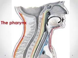
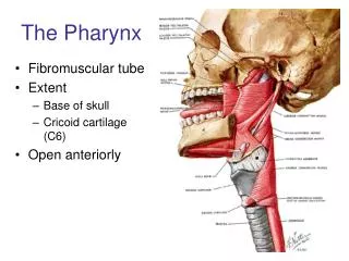
PHARYNX. Extent: base of the skull to C6 vertebra Function: Channel for food Air through NP Air & food through OP Measurements: Length: 12 to 14cms . Width: nasopharynx (3.5cm) pharyngo-oesophageal junction (1.5cm). C1. C2. C3. C4. C5. C6. Boundaries:. Superiorly:

E N D
Extent: • base of the skull to C6 vertebra Function: • Channel for food • Air through NP • Air & food through OP Measurements: • Length: 12 to 14cms. • Width: nasopharynx (3.5cm) pharyngo-oesophageal junction (1.5cm).
C1 C2 C3 C4 C5 C6 Boundaries: • Superiorly: • Inferiorly: • Posteriorly: • Anteriorly: • On each side:
In front: • Communicates with nasal cavity through choanae • Communicates with oral cavity through oropharnygeal isthmus • Communicates with larynx through laryngeal inlet • Behind: • Upper six cervical vertebrae and intervertebraldiscs,pre & para vertebral muscles, retropharyngeal space • On either side: styloid process, carotid sheath,lateral lobe of thyroid gland
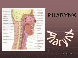
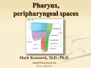
Parts: • Nasopharynxbehind the nose. • Oropharynx behind the oral cavity. • Laryngopharynxbehind the larynx.
Parts • Nasopharynx • Oropharynx • Laryngopharynx
Nasopharynx • situated behind the nasal cavity and above the soft palate
Boundaries • Anterior wall : Communicates with nasal cavity through choanae. • Posterior wall (roof): Body of the sphenoid, basiocciput, anterior arch of atlas.
Nasopharynx: Roof and posterior wall: It shows pharyngeal or nasopharyngeal tonsil.
Lateral wall presents the following features: • Pharyngeal opening of auditory tube • Tubal elevation • Salpingopharyngeal fold • Levator veli palatini • Pharyngeal recess (fossa of Rosenmuller)
Nasopharyngeal tonsil • Formed by aggregation of lymphoid tissue beneath the mucous membrane. • Present in the internal aspect of the roof & posterior wall. • More prominent in children and atrophies in adults. • when enlarged due to infection, is known asadenoid which obstructs the nasal respiration.
Pharyngeal isthmus • Formed from the fibers of palatopharyngeus with upper fibers of superior constrictor muscle. • Closed during swallowing or blowing air through mouth
Oropharynx: • Extension: • Superiorly: • Anteriorly: • Inferiorly: • Posteriorly: related to axis & C3 vertebra • Lateral wall presents tonsillar fossa lodging palatine tonsil
Oro pharynx • Lies behind the oral cavity • In front : • communicates in front with the oral cavity through the oropharyngeal isthmus • Boundaries of oropharyngeal isthmus • Above: Soft palate • Below: Dorsum of the tongue • On each side:Palatoglossal arch
Behind : by the bodies of C2,C3 vertebrae • Below : communicates with the laryngopharynx • Lateral wall: bears tonsillarfossa that lodges palatine tonsils C2 C3
Tonsillar fossa: • Anteriorly,palatoglossal arch • Posteriorly, palatopharyngeal arch • Lodges the Palatine tonsil.
Laryngopharynx: • Extension: • Anterior wall: has inlet of larynx • Posterior wall: related to C3,C4,C5 & C6 vertebra • Lateral wall presents piriform fossa
Inlet of larynx Aryepiglottic fold Piriform fossa Piriform fossa • Present in the lateral wall of laryngopharynx • Boundaries: • Medial: Aryepiglottic fold • Lateral: thyrohyoid membrane & thyroid cartilage • Relation: • Floor is related to internal laryngeal nerve • Importance: • It usually lodges foreign bodies like fish bones. Any attempt to remove these may damage the internal laryngeal nerve which leads to anaesthesia of upper part of the larynx.
Note • Thyrohyoid membrane is pierced by the internal laryngeal nerve & superior laryngeal vessels • Waldeyer’s ring: • Ringof lymphoid tissue encircling the cephalic part of the air and food passage • Includes lingual, palatine,tubal, nasopharyngeal tonsil • Helps in primary defense .
Structure of the pharynx: Outside to inside: • Areolar coat(buccopharyngeal fascia) • covers the outer surface of the constrictor muscles • Attached above to the base of skull & forms the anterior boundary of retropharyngeal space • Not well defined • Muscular coat: consists of outer circular & inner longitudinal muscle coat. • Submucosal layer: pharyngobasilar fascia lies between the submucosa and muscular layer • Mucosal layer