Download
1 / 32
330 likes | 528 Views
Oral cavity, pharynx. Dr. L. Kiss Anna Department of Anatomy , Histology and Embryology Semmelweis University 2019. Parts of the body. Regions. Th12/L1. Regions. Supra-and infrahyoid muscles. Cervical trigone. Submental trigone. Submandibular trigone. Carotid trigone.

E N D
Oral cavity, pharynx Dr. L. Kiss Anna Department of Anatomy, Histology and Embryology Semmelweis University 2019.
Regions Th12/L1
Cervicaltrigone Submentaltrigone Submandibulartrigone Carotidtrigone Omoclaviculartrigone
Cervicaltrigone Submentaltrigone: lymphnodes Submandibulartrigone: submandibulargland lateralgroove of thetongue (nerves) Carotidtrigone: carotidsheath (commoncarotidartery internaljugularvein vagusnerve) Supraclaviculartigone: 2 parts: omotrapezoidtrigone: cervicalplexus omoclaviculertrigone: brachialplexus subclavian art. apex of thelung
Cervicaltrigones omoclaviculartrigone: brachialplexus subclavial art. and vein. apex of thelung carotidtrigone
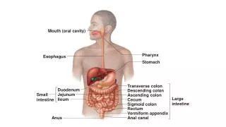
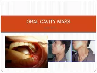
Oralcavity: Anteriorborder: lips Roof: hard and softpalate Floor: mylohyoidmuscle Lateralborder: cheeks (buccinatormuscle) Isthmusfaucium: exittowardthepharynx 2 parts: oralcavityproper+vestibulum Vestibulum: slitlikespacebetweenthelips, cheeks and teeth Oralcavityproper: tongue teeth salivaryglands
arcus palatoglossus uvula arcus palatopharyngeus * * *: tonsilla palatina tongue
hard palate soft palate uvula
Teeth dentes permanentes dentesdecidui dentes permanentes
canine molars incisive praemolars Dentespermanentes
crown (coronadentis) neck gingiva root (radix) pulp (pulpa) bone
enamel dentine cementum periodontum
Innervation of the teeth Trigeminal ganglion Maxillary nerve Inf. alveolar nerve (mandibular n.)
Tongue root sulcusterminalis palatine tonsil palatine tonsil lingual tonsils circumvallate papillae foliate papillae apex fungiform papillae filiform papillae
Taste sensation green: bitter blue: sour red: salty orange: sweet
Innervation of the tongue Sensory innervation: general: lingual n. (V./3) – anterior 2/3 glossopharyngeal n. (IX.) – posterior 1/3 vagus n. (X.) – the most posterior part taste: chorda tympani (VII.) – anterior 2/3 glossopharyngeal n.(IX.) – posterior 1/3 Motory innervation: hypoglossal n. (XII.)
Salivary glands I. parotis parotid duct Opens: vestibule, upperrow of theteeth (2nd molar)
Salivary glands II. parotis submandibular gland
Sublingual region frenulum of the tongue sublingual gland
Salivary glands III. sublingual gland mandible root of the tongue duct of the submandibular gland submandibular gland
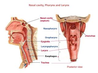
Pharynx nasopharynx tongue oropharynx soft palate laryngopharynx epiglottis
Waldeyer’s lymphatic ring Tonsils present in the ishtmus faucium: • tonsilla pharyngea • tonsillae tubariae • tonsilla palatina • tonsilla lingualis
Muscles of thepharynx: elevators stylopharyngeal m. salpingopharyngeal m. palatopharyngeal m.
Pharynx uvula root of the tongue piriform recess epiglottis
Esophagus 1.) p. cervicalis: 6. cervical vertebra, lower surface of the cricoid cartilage 2.) p. thoracalis: post. mediastinum hiatus esophageus (10. thoracal vertebra) 3.) p. abdominalis
Esophagus Physiological narrowing of the esophagus: A.) 15 cm from the teeth row: pharynx-esophagus transition – cricoid cartilage B.) 24 cm from the teeth row: aortic arch, left principal bronchus C.) 40 cm from the teeth row: at the cardia (sphincter)