G.I. Bleeding
G.I. Bleeding. Presented by: Ahmed T. Al-Suwaidi Mohamed S. Al-Hoqani. G.I. Bleeding Case. 50 yrs, Pakistani, male C/O: Bleeding/rectum & Abd. pain Painless bleeding, 1 yr – excess bleeding, 1 month Black, 4-5 times/day, little quant. Abd. pain Vomiting, 1 week. G.I. Bleeding Case.

G.I. Bleeding
E N D
Presentation Transcript
G.I. Bleeding Presented by: Ahmed T. Al-Suwaidi Mohamed S. Al-Hoqani
G.I. Bleeding Case • 50 yrs, Pakistani, male • C/O: Bleeding/rectum & Abd. pain • Painless bleeding, 1 yr – excess bleeding, 1 month • Black, 4-5 times/day, little quant. • Abd. pain • Vomiting, 1 week
G.I. Bleeding Case • M.H: * no peptic ulcer disease * no medications (NSAIDs) * no urinary symptoms * not known DM, HPTN, IHD ** weight loss
G.I. Bleeding Case • O/E: * Afebrile * no pallor * not dyspneaic * no lymphoadenopathies * no S.C.L.N
G.I. Bleeding Case • Vital Signs: * Pulse: 78 bts/min * BP: 130/80 * RR: 18 br/min • Heart: NAD • Lung: NAD
G.I. Bleeding Case • Abd.: * not distended * no epigast. tenderness * tender, firm, partly mobile mass at Rt lumbar region. * spleen not palpable * Lt lobe liver palpable, mildly tender * bowel sounds present
G.I. Bleeding Case • PR: * no enlarged piles * no active bleeding * no palpable mass * no blood on finger • ECG, CBC, Sr Amylase, Bleeding profile, Abd X-ray, fecal loading ascending colon
G.I. Bleeding Case • Lab Results: * Hb: 14.1 g/dl * Plt: 252 * 103 * Hypochromic, microcytic * PT: 17.3 sec * aPTT: 35.4 sec * Sr Amy: 129 U/l 106 U/l * Na+: 140 mmol/l * K+: 4.1 mmol/l * BUN: 17 mg/dl
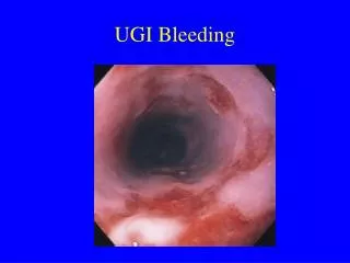
G.I. Bleeding • Acute Vs Chronic • Acute Upper G.I.Bleeding: • Acute Lower G.I.Bleeding:
Acute Upper G.I. Bleeding • Haematemesis • Melaena • Site & Time
Acute U.G.I. Bleeding • ·Aetiology: • 1. Drugs (Aspirin & NSAIDs) • 2. Alcohol • 3.Chronic peptic ulceration (50% of GI hemorrhage) • 4.Others: reflux esophagitis, varices, gastric carcinoma, acute gastric ulcers & erosions.
Acute U.G.I. Bleeding • ·Clinical approach: • 1. recent (24 hrs), then hospitalized. • 2. if small amount, no immediate Tx, because CVS can compensate • 3. 85% stop bleeding during 48 hrs • 4. history helps in diagnosing the cause of the hemorrhage, eg: long history of indigestion, or previous hem. from ulcers.
Acute U.G.I. Bleeding ·Clinical approach: • 5. factors include: ·age (60 +) • ·amount of bld lost • ·continuing visible bld loss. • ·signs of chronic liver disease • ·classical clinical features of shock
Acute U.G.I. Bleeding ·Clinical approach: 6. liver disease severe, recurrent bleeding (if from varices) 7. splenomegaly portal hypertension
Acute U.G.I. Bleeding • ·Immediate management: ** Emergency management: • ·History + exam. • ·Monitor: pulse & BP /30 min • ·Bld sample: haemoglobin, urea, electrolytes, grouping & cross-matching • ·I.v. access
Acute U.G.I. Bleeding ** Emergency management (cntd): • · Bld transfusion in case of • 1) shock 2) haemoglobin <10 g/dl • ·Urgent endoscopy • ·Surgery when recommended
Acute U.G.I. Bleeding **Shock management: • ·ABC • · Airway: endotracheal tube, oropharyngeal airway. *Give oxygen
Acute U.G.I. Bleeding **Shock management (cntd): • ·Breathing: support respiratory function * Monitor: resp. rate, bld gases, chest radiograph • · Circulation: expand circulating volume: blood, colloids, crystalloids support CVS function: vasodilators * Monitor: skin color, peripheral temp., urine flow, BP, ECG
Acute U.G.I. Bleeding • ·General Investigations: 1. Hb, PCV 2. CBC (WBC … etc) 3. Bld glucose 4. Platelets, coagulation 5. Urea, creatinine, electrolytes 6. Liver biochem. 7. Acid-base state 8. Imaging: chest & abd. radiography, US, CT
Acute U.G.I. Bleeding **General management: • ·Blood volume 1. restore volume to normal 2. transfusion • ·Endoscopy 1. shock, suspected liver disease or continued bleeding 2. control varices or ulcers to reduce re-bleeding
Acute U.G.I. Bleeding **General management: • ·Drug therapy 1. H2 – receptor antagonists 2. proton pump inhibitors • ·Factors in reassessment 1. age: 60 + greater mortality 2. recurrent hemorrhage: +++ mortality 3. re-bleeding: mostly within the 1st 48 hrs 4. surgical procedures in case of severe bleeding.
Lower gastrointestinal haemorrhage Causes • Diverticular disease • Angiodysplasia • Inflammatory bowel disease • Ischaemic colitis • Infective colitis • Colorectal carcinoma
Investigation • Mostpatients are stable and can be investigated once bleeding has stopped • In the actively bleeding patient consider • Colonoscopy - can be difficult • Selective mesenteric angiography • Requires continued bleeding of >1 ml/minute • May show angiodysplastic lesions even once bleeding has ceased
Radionuclide scanning • Uses technetium-99m labeled red blood cells
Management • Acute bleeding tends to be self limiting • Consider selective mesenteric embolisation if life threatening haemorrhage • If bleeding persists perform endoscopy to exclude upper GI cause • Proceed to laparotomy and consider on-table lavage an panendoscopy • If right-sided angiodysplasia perform a right hemicolectomy • If bleeding diverticular disease perform a sigmoid colectomy • If source of colonic bleeding unclear perform a subtotal colectomy and end-ileostomy