Download
1 / 42
420 likes | 543 Views
The respiratory system is essential for life, comprising the lungs and air passages, responsible for oxygen intake and carbon dioxide removal. Key components include the nose, pharynx, larynx, trachea, bronchi, bronchioles, and alveoli. This system must function continuously, providing a brief supply of oxygen. The process of ventilation involves two phases: inhalation and exhalation, regulated by the diaphragm and intercostal muscles. Gas exchange occurs in the alveoli, where oxygen enters the bloodstream and carbon dioxide is expelled, ensuring cell health and function.

E N D
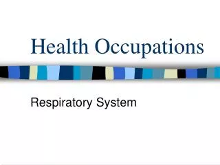
Health Occupations Respiratory System
Respiratory System • Consists of • Lungs • Air passages • Responsible for • Taking in oxygen needed by ALL body cells • Removing carbon dioxide & waste produced by cells • 4-6 minute supply of oxygen • System MUST work continuously
Parts of the Respiratory System • Nose A. Air enter via 2 nostrils B. Nasal septum – divides nose into 2 hollow nasal cavities C. Lined with mucous membranes with a rich blood supply D. Air enters cavity 1. Warmed 2. Filtered 3. Moistened
E. Mucous is produced by mucous membranes 1. Moistens air 2. Traps pathogens & dirt F. Cilia – helps to move mucous 1. Pushes trapped particles towards esophagus where it is swallowed G. Olfactory receptors – sense of smell H. Nasolacrimal ducts – drain tears from eyes to nose, provides extra moistures
2. Sinuses • Cavities in skull surrounding nasal area • Connected to nasal cavity by short ducts • Lined with mucous membranes to warm & moisten air • Provides resonance for voice
3. Pharynx • Throat, lies directly behind nasal cavities • Air leaves nose & enters pharynx • 3 sections • Nasopharynx • Upper portion, behind nasal cavities • Contains adenoids • Contains Eustachion tube (to middle ear)
2. Oropharynx a. Middle section, behind mouth b. Receives air from nasopharynx c. Receives food & air from mouth 3. Laryngopharynx a. Bottom section of pharynx b. Contains esophagus, carrying food to stomach c. Contains trachea, carrying air to & from the lungs
4. Larynx • Voice box, between pharynx & trachea • Nine layers of cartilage • Thyroid cartilage – Adam’s apple is largest • Vocal cords – 2 folds in larynx • Glottis – opening between the vocal cords • As air leaves lungs, vocal cords vibrate & produce sound. Tongue & lips act on sound to produce speech • Epiglottis – leaflike piece of cartilage that closes the opening of the larynx when swallowing to prevent food & liquid from entering the respiratory tract
5. Trachea • Windpipe, tube extending from larynx to the center of the chest • Carries air between pharynx & bronchi • C-shaped cartilage, in a series, open on the dorsal surface, to keep trachea open
6. Bronchi • Trachea divides into 2 of these near the center of the chest • Right bronchus – shorter, wider, more vertical • Left bronchus – longer, narrower, less vertical • Each enters a lung, carries air from trachea to lung • In lung, bronchi divide into smaller & smaller bronchi • Lined with cilia & sticky mucous (phlegm) to catch dust & germs • Produces 125 mL of mucous/day that is removed by cilia
7. Bronchioles • Very small bronchi • Terminal bronchioles – smallest • End in sacks called alveoli
8. Alveoli • Look like a bunch of grapes • Adult lung – 500 million alveolis • Consists of 1 layers of squamous epithelial tissue & has many capillaries • Capillaries allow oxygen & carbon dioxide exchange between blood & lung • Inner surface of alveoli are covered with a fatty substances (surfactant) that prevents them from collapsing
9. Lungs • Organs of respiratory system, contain the bronchi & alveoli • Right lung 1. 3 sections or lobes • Left lung 1. 2 lobes, smaller because of heart’s location • Pleura • Sac or membrane containing lungs • 2 layers • Visceral pleura – attaches to lung surface • Parietal pleura – attaches to chest wall
E. Pleura space 1. Located between 2 layers 2. Filled with pleural fluid 3. Lubricates the membranes, prevents friction as lungs expand during breathing F. Located in thoracic cavity along with heart & blood vessels G. Mediastinum – space separating the lungs & contains esophagus, heart, & bronchi
Ventilation • Process of breathing • 2 phases • Inspiration or Inhalation • Breathing in air • Diaphragm (dome shaped muscles between the thoracic & abdominal cavities) & intercostal muscles contract & enlarge the thoracic cavity to create a vacuum (moves downward) • Air rushes through airways to alveoli • Gas exchange takes place in alveoli • Process is called respiration
3. Expiration or exhalation • Diaphragm & intercostal muscles relax • Air is forced out of lungs & air passages
4. Respiration controlled • Respiratory center in the medulla oblongata of brain • Respirations increase due to • Increased CO2 in blood • Decreased O2 in blood (asthma, COPD, CHF) • Mostly involuntary, but person can control rate
Stages of respiration • External respiration – exchange of O2 & CO2 between LUNGS & BLOODSTREAM • Oxygen enters alveoli • Oxygen concentration in alveoli is higher than concentration in blood capillaries • Oxygen leaves alveoli to enter capillaries • CO2 goes the reverse way, leaving the capillaries to enter the alveoli (because CO2 is higher in the capillaries than the alveoli) • CO2 expelled during exhalation
2. Internal respiration • Exchange of CO2 & O2 between BLOOD & TISSUE CELLS • O2 carried to tissue cells by blood • Concentration of O2 is higher in blood, so it leaves blood for tissues • Cells use O2 & nutrients to produce energy, water, & CO2 • CO2 concentration is higher in tissues than blood, so CO2 leaves tissues to enter blood
3. Cellular respiration • Tied in with internal respiration • Cells use O2 & nutrients • This process produces energy, water, & CO2 Once CO2 is transported back to bloodstream (internal respiration) it enters the lungs & is expelled by exhalation (external respiration)
Assessment • Rate • Breaths per minute • Varies with age, posture, exercise, temperature, etc • Children have faster rate than adults • Normal adult 14 – 20 per minute • Eupnea – normal respiration • Dyspnea – difficult or labored respiration • Tachypnea – respirations > 24 per minute • Bradypnea – respirations < 10 per minute
2. Character • Rhythm of respiration • Should have regular rhythm • Apnea – absence of respirations • Cheyne-Stokes respirations – abnormal pattern of respiration, labored, followed by apnea • May be dry, normal, wet, or shallow
3. Sounds • Breath sounds – heard with stethescope • All should be clear & dry • Wheezing – high pitched • Rales – like hair rubbing together • Rhonchi – wet bubbly noises
4. Lung volumes • Respiratory capacity – amount of air that can be brought into lungs • Measured with a spirometer • Depends on age & physical condition • Lung volume – measure of respiratory capacity • Tidal volume – amount of air normally exchanged with each inspiration & expiration • Inspiratory reserve volume – additional amount of air that can be inhaled with a conscious effort
Expiratory reserve volume – additional amount of air that can be exhaled with a conscious effort • Vital capacity = TV + IR + ER • Residual volume – amount of air always in lungs to maintain shape • TLC (Total lung capacity) = TV + IR + ER + RV
TOTAL LUNG Capacity VITAL CAPCITY Tidal volume Inspiratory Reserve Volume Expiratory Reserve Volume Residual Volume
5. Blood gases • Measure amount of gases in blood and blood pH • Accurate assessment • Hypoxia – not enough oxygen in blood
Abnormal conditions • Asthma – respiratory disorder usually caused by sensitivity to allergen, dust, pollen, animal, or food. Can be caused by stress, overexertion, & infection • Bronchospasms narrow opening of bronchioles • Mucous production increases • Edema develops in mucosal lining • Symptoms – dyspnea, wheezing, coughing, sputum, chest tightness • Treatment – bronchodilators, anti-inflammatory, epinephrine, O2, eliminate allergens
2. Bronchitis • Inflammation of bronchi & bronchioles • Acute bronchitis – caused by infection • Symptoms – productive cough, dyspnea, chest pain, fever • Treatment – antibiotics, expectorants, rest, FF • Chronic bronchitis – results from frequent attacks of acute bronchitis & long term exposure to smoking or pollutants • Chronic inflammation, damaged cilia, enlarged mucous glands • Symptoms –excessive mucous with productive cough, wheezing, dyspnea, CP, prolonged air expiration • No cure, may use antibiotics, bronchodilators, respiratory therapy
3. COPD • Chronic lung disease resulting in obstruction of airways • Caused by chronic asthma, chronic bronchitis, emphysema, TB, smoking, allergies, & chronic respiratory infections • See symptoms of chronic bronchitis
4. Emphysema • Noninfectious chronic respiratory condition occurring when walls of alveoli deteriorate & lose elasticity • CO2 remains trapped in alveoli & there is poor gas exchange • Causes – heavy smoking, exposure to pollutants • Symptoms – dyspnea, feeling of suffocation, pain, barrel chest, chronic cough, cyanosis, rapid respirations with prolonged expiration, eventual respiratory failure & death • No cure, treat with bronchodilators, prompt tx of respiratory infections, O2, resp tx, avoid smoking
5. Epistaxis • Nosebleeds • Occur when capillaries in nose become congested & bleed • Causes – nose injury, HTN, chronic infection, anticoagulants, blood diseases (hemophilia, leukemia) • Treatment – pinch nostrils towards septum, tilt head slightly forward, cold compresses. Sometimes necessary to insert nasal packs or cauterize
6. Influenza • Flu • Highly contagious, viral infection of upper respiratory system • Sudden onset, chills, fever, cough, sore throat, runny nose, muscle pain, fatigue • Treatment – bedrest, fluids, analgesics, Tylenol • Immunizations recommended for elderly, people with chronic disease, pregnant, health care workers, children
7. Laryngitis • Inflammation of larynx & vocal cords • Frequently occurs with other respiratory infections • Symptoms – hoarseness, loss of voice, sore throat, dysphagia • Treatment – rest, limited voice use, fluids, meds if infection
8. Lung cancer • Leading cause of cancer deaths • Preventable disease – main cause is tobacco exposure • 3 types of lung CA – Squamous cell, small cell, adenocarcinoma • Early stages – no symptoms • Later stages – chronic cough, hemoptysis, dyspnea, fatigue, weight loss, CP • Prognosis poor because of late dx • Treatment – surgical removal of cancerous part of lung, radiation, chemo
9. Pleurisy • Inflammation of pleura • Usually in conjunction with other infections • Symptoms – sharp stabbing pain when breathing, crepitus, dyspnea, fever • Treatment – rest, meds to relieve pain & inflammation, serious cases - thoracentesis
10. Pneumonia • Inflammation or infection of lungs with exudates in alveoli (fluid) • Usually caused by bacteria, viruses, or chemicals • Symptoms – chills, fever, CP, productive cough, dyspnea, fatigue • Treatment – bedrest, fluids, antibiotics, resp tx, pain meds
11. Rhinitis • Inflammation of nasal mucous membrane • Symptoms – runny nose, soreness, congestion • Causes – infections, allergens • Treatment – fluids, meds to relieve congestion
12. Sinusitis • Inflammation of mucous membranes in sinuses • May involve 1 or more sinuses • Usually caused by virus or bacteria • Symptoms – HA or pressure, thick nasal discharge, congestion, loss of voice resonance • Treatment – analgesics, antibiotics, decongestants, moist inhalations, sometimes surgery if sinusitis is chronic
13. Tuberculosis • Infectious lung disease caused by mycobacterium tuberculosis • WBC surround invading TB organisms & wall them off in nodules called tubercules in lungs • TB organisms can remain dormant in tubercules but can become active TB later if resistance is lowered • Symptoms of active TB – fatigue, fever, night sweats, weight loss, hemoptysis, CP • Treatment – drugs for 1or more years, good nutrition, rest • New strain of TB is drug resistant
14. Upper respiratory infection • URI, common cold • Inflammation of mucous membranes lining upper respiratory tract • Caused by viruses, very contagious • Symptoms – runny nose, fever, watery eyes, congestion, sore throat, hacking cough • No cure, symptoms last about 1 week • Treatment – analgesics, Tylenol, rest, FF, antihistamines to relieve symptoms
15. Cystic fibrosis • Genetic disease of exocrine glands in which the mucous in respiratory system becomes thick • Excessive salt also forms on skin • Also have digestive difficulties • Need to give intensive pulmonary care • Outlook is better now