Download
1 / 42
420 likes | 517 Views
Explore often overlooked atraumatic conditions affecting SC joint & their diagnostic features. Includes details on Friedrich's Dz, SC hyperostosis, OA, subluxation, and septic arthritis.

E N D
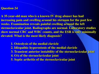
Question 24 A 35-year-old man who is a known IV drug abuser has had increasing pain and swelling around his sternum for the past few weeks. Examination reveals painful swelling around the left sternoclavicular joint. Radiographs are normal. Laboratory studies show normal CBC and WBC counts, and the ESR is only minimally elevated. What is the most likely diagnosis? 1. Osteolysis of the medial clavicle2. Idiopathic hyperostosis of the medial clavicle 3. Transient anterior instability of the sternoclavicular joint 4. OA of the sternoclavicular joint 5. Septic arthritis of the sternoclavicular joint
Answer: 5 Clarke Several atraumatic conditions that may affect the SC joint are often overlooked or misdiagnosed. Friedrich’s Dz (Aseptic necrosis of medial clavicle) – irreg of the SC jt and bony defect and destruction of the medial clavicle. Unilateral, women SC hyperostosis (SCCH) – bone overgrowth and soft-tissue ossification of the medial clavicle, upper ribs, and sternum. Etiology unknown. A/w aseptic-pustular lesions of the palms and soles. OA of SC jt – narrowing of the jt space, osteophytes, subchondral sclerosis, and cysts on both sides of the jt. Most wear/degen occurs in inferior part of head medial clavicle. Spont. Subluxation SC jt – teenagers, palpable and visual anterosuperior subluxation of the medial end of clavicle when the arm is raised to the overhead position. Spont reduces when arm is brought down. No h/o trauma. Not painful. Septic Arthritis – Spontaneous swelling with joint subluxation. Erosive changes of the medial clavicle and manubrium with some fragmentation and sclerosis. CT aids dx. Predisposing conditions include IVDA and HIV+. Gram Neg bacteria in IVDA Hiramuro-Shoji et al. Atraumatic conditions of the SC joint. J Shoulder Elbow Surg 2003;12:79-88. Rockwood et al. Disorders of the SC joint. The Shoulder, ed 2. Philadelphia, PA, Lippincott Williams and Wilkins, 1998:555-609.
Question 36 A 79-year-old man with a long standing rotator cuff tear reports that the steriod injections he has been receiving no longer provide pain relief. A radiograph is shown in Figure 7. Treatment should consist of 1- primary rotator cuff repair. 2- rotator cuff repair with an orthobiologic patch 3- latissimus dorsi transfer 4- shoulder arthrodesis 5- humeral head arthroplasty
Question 36 answer: 5 T. Wimmer The radiograph is an AP right shoulder that shows superior migration of the humeral head with severe glenohumeral degenerative changes and erosive changes at the AC joint and of the acromion. These are classic findings of the so called “cuff tear arthropathy”. Once the function of the rotator cuff is lost through a massive tear, often involving all of the supraspinatus and infraspinatus, this pattern of shoulder arthritis may develop. The rotator cuff is an important dynamic stabilizer of the glenohumeral joint acting to centralize the humeral head on the glenoid. It also forms a force couple with the deltoid for active shoulder motion. Loss of its function leads to superior migration of the humeral head, unopposed action of the deltoid, eccentric loads being applied to the humeral head and gleniod, and significant disruption of joint biomechanics.Treatment of this entity, significant glenohumeral arthritis in association with rotator cuff deficiency, is more complicated than treatment of arthritis without cuff deficiency.
Answer: 5 T. Wimmer Often these patients are treated with non-operative measures including injections as long as they are capable of satisfactory function with acceptable comfort relief. Indication for surgery is usually intractable pain unresposive to conservative care. Requirements for surgery are: 1. Intact deltoid function and 2. Intact coracoacromial arch. Total shoulder arthroplasty has resulted in early glenoid loosening at an unacceptable rate. This is likely secondary to eccentric loading of the glenoid component by a high riding humeral head. The treatment of choice in this setting is hemiarthroplasty. This provides excellent pain relief and return of function. Collins’ article is a review of arthroplasty for arthritis and rotator cuff deficiency. They agree hemiarthroplasty is the treatment of choice and describe operative techniques. Zuckerman’s article is a retrospective review of 15 cases of hemi for cuff tear arthropathy.
References T. Wimmer Collins DN, Harryman DT II: Arthroplasty for arthritis and rotator cuff deficiency. Orthop Clin North Am 1997;28:255-239. Zukerman JD, Scott AJ, Gallagher MA: Hemiarthroplasty for cuff tear arthropathy. J Shoulder Elbow Surg 2000;9:169-172.
Joe CarneyQuestion 40A 24 year old baseball pitcher who has had shoulder pain for the past 6 months undergoes glenohumeral arthroscopy. The anatomic finding shown in Figure 11 reveals a 1) Bankart lesion2) Superior labral tear3) Rotator cuff tear4) Normal anatomic variant5) detached middle glenohumeral ligament
H S MGL RI L G
Answer: normal anatomic variant The figure has been labeled for reference: H=Humeral articular surface G=Glenoid L=Labrum MGL=Middle Glenohumeral ligament S=Subscapularis RI=Rotator Interval There is a wide variability in the anatomy of the anterosuperior portion of the labrum anteriorly between the origin of the long head of the biceps and the equator of the glenoid. Rao et al and Williams et al found the overall prevalence of anterosuperior labral variation was 13%.
Answer: normal anatomic variant One such described normal variant is the presence of a sublabral foramen, or sulcus, between the normally developed anterosuperior portion of the labrum and the glenoid articular cartilage with a sheet like middle glenohumeral ligament attaching to the labrum (seen in the photograph). This variant is different from the “Buford Complex” normal variant which Williams et al described as an absence of labral tissue at the anterosuperior portion of the labrum and a cord-like middle glenohumeral ligament that attaches to the superior part of the labrum at the base of the biceps tendon. It is widely accepted that anatomical variants in the anterosuperior quadrant of the labrum do not contribute to laxity or instability and therefore should not be addressed surgically as part of an operation to treat instability. Repair of the normal variants has been reported to produce pain and restricted range of motion.
Question 51 An 18-year-old collegiate swimmer has had bilateral shoulder pain for the past 6 months. She also notes frequent episodes of numbness throughout her dominant hand and prominent scapular winging on one side. Based on these signs and symptoms, examination will most likely reveal 1. clinodactyly. 2. negative ulnar variance. 3. absent distal palmar crease. 4. hypermobile patellae. 5. tarsal coalition.
Answer: 4M. RobinsonMultidirectional instability patients commonly present with shoulder pain (often bilateral) and transient neurological symptoms. Neurological symptoms include numbness, tingling, and weakness. MDI is associated with generalized ligamentous laxity. Generalized ligamentous laxity signs include elbow hyperextension, metacarpophalangeal joint hyperextension, genu recurvatum, and patellar subluxation. Schenk TJ, Brems JJ: Multidirectional instability of the shoulder: Pathophysiology, diagnosis, and management. JAAOS 1998;6:65-72.Neer CS II, Foster CR: Inferior capsular shift for involuntary inferior and multidirectional instability of the shoulder: A preliminary report. JBJS 2001;83:1586.
Question 66 A 65-year old woman who underwent total shoulder arthroplasty on the left side 1 year ago now reports that she is unable to tuck in her shirt behind her back using the left hand. Examination reveals weakness with a belly-press test on the left side. What is the most likely diagnosis? Excessive glenoid retroversion Excessive humeral head retroversion Inadequate postoperative therapy Subscapularis insufficiency 5. Axillary nerve injury with deltoid dysfunction
Q 66 preferred response 4 Jim Harris The physical exam described in the question gives the answer. Weakness inbelly-press is associated with subscapularis dysfunction. Miller in her cited article reports 67.5% of TSR patients(27/32) had reduced subscap function by lift-off and belly-press. Of those 27, 92% were unable to tuck in a shirt. All other answers are know complications of TSR but subscap dysfunction is the most common. Tokish, in another article in J Shoulder Elbow Surg 2003, proved via EMG that “belly-press” isolates upper subscap better than lift-off and vice versa for lower subscap. Miller J Shoulder Elbow Surg 2003 Colfield Instr Course Lect 1990
Question 89 What structure in the shoulder is the primary restraint to anterior dislocation at 90deg of abduction?1- Inferior labrum2- Superior glenohumeral ligament3- Middle glenohumeral ligament4- Posterior band of the inferior glenohumeral ligament5- Anterior band of the inferior glenohumeral ligament
Answer: 5 DTSchroderBurra and Andrews: SGHL is the primary stabilzer against inferior translation when the arm is at the side. When the arm is abducted beyond 45deg, the IGHL is the primary static stabilizer against inferior translation. The IGHL is the primary static stabilizer of the abducted arm. The anterior band is the primary restraint to anterior dislocation at 90deg abduction . In the midrange of abduction the middle glenohumeral ligament is the primary stabilizer to anterior translation. The posterior band of the IGHL and the rotator interval capsule are the primary static posterior stabilizers.The rotator cuff is the primary dynamic stabilizer with the subscapularis resisting anterior translation. O’Brien et al.: A cadaveric study of 11 shoulders. Arthroscopic exam revealed a prominent anterior and posterior band of the IGHL, present in all shoulders with little variability (contrary to variable appearance of MGHL and SGHL). With the arm in 90 deg of abduction and external rotation, the anterior band and interposed axillary pouch fan out and become taut. With internal rotation and 90 deg of abduction the posterior band of the IGHL and interposed pouch fan out and become taut.Burra G, Andrews JR: Acute shoulder and elbow dislocations in the athlete. Orthop Clin North Am 2002;33:479-495.O’Brien SJ, Neves MC, Arnoczky SP, et al: The anatomy and histology of the inferior glenohumeral ligament complex of the shoulder. Am J Sports Med 1990;18:449-456.
102. A 15-year-old girl who has been swimming competitively for many years reports increasing pain in her shoulder during the backstroke and unremitting numbness in her little and ring fingers. She also notes a prominence of her shoulder blade on the affected shoulder. Examination will most likely reveal which of the following findings? 1 – Positive O’Brien’s test 2 – Positive lift-off test 3 – Limited internal rotation 4 – Deltoid weakness 5 – Serratus anterior weakness
Answer 5 Carter Maurer A swimmer with many years of increasing pain likely has some degree of multidirectional instability. The articles referenced state that scapular dyskinesis can be caused by any number of potential internal derangements of the shoulder (including MDI) and presents clinically by prominence of the medial boarder of the scapula due to serratus anterior weakness. The answer can also be obtained directly by knowing that serratus anterior weakness causes prominence of the scapula. This can be demonstrated with a wall push-up. There is no indication of labral pathology which would be demonstrated with O’Brien’s test (pain with thumb down but not with palm up). A swimmer with MDI would likely have increased ROM not limited. There is no indication of deltoid weakness in the question. Kibler WB, McMullen J: Scapular dyskinesis and its relation to shoulder pain. J Am Acad Orthop Surg 2003;11:142-151.Kibler WB, Uhl TL, Maddux JW, Brooks PV, Zeller B, McMullen J: Qualitative clinical evaluation of scapular dysfunction: A reliability study. J Shoulder Elbow Surg 2002;11:550-556
Question 124 - A 50 year-old recreational athlete has had severe pain and loss of motion in his right nondominant shoulder for the last 12 months. History reveals that he underwent surgery for recurrent dislocations 22 years ago. A radiograph is shown in Figure-35. Management should consist of: 1. shoulder fusion. 2. arthroscopic debridement. 3. resection arthroplasty. 4. total shoulder arthroplasty. 5. manipulation under anesthesia.
Answer: 4 KA Menzel Total shoulder Arthroplasty. The history and exam findings in this patient support what has been termed “arthritis of dislocation.” Typically the patient is younger and has had one or more previous surgeries for shoulder instability. Over time they develop increasing pain and loss of motion. Radiographs show progressive glenohumeral arthrosis and decreased joint spacing with osteophyte formation. The etiology of the arthritis in these patients seems to be multifactorial. All patients have a history of a single or repeated episodes of dislocation or subluxation. Although some data show no correlation with the number of episodes of instablilty and the development of arthritis, the pattern of instability does play a role in the development of future arthritis. Several factors relating to surgical treatment of instability also seem to play a role. These include: overtightening the anterior structures, substantially limiting external rotation and increasing joint-loading posteriorly; misdiagnosing the degree and direction of instability leading to insufficient surgical correction; migrated or improperly placed surgical metal implants; technical errors (eg-gouging cartilage with arthroscope); along with others.
Answer: 4 (cont.) KA Menzel The literature has shown success in treating these patients with hemiarthroplasty/total shoulder arthroplasty depending on the degree of glenoid involvement. In these patients there is no role for arthroscopic debridement or MUA as neither will treat the arthritis nor return the patient to full painless motion. Shoulder arthrodesis should be reserved for those patients with a history of septic arthritis and painful paralysis about the shoulder, and is not indicated for aseptic arthritis regardless of the patient’s age. The patient in the question has radiograhs which show significant arthritic changes to both sides of the glenohumeral joint; therefore, the correct treatment option is total shoulder arthroplasty.-Bigliani LU, Weinstein DM, Glasgow MT, Pollock RG, Flatow EL: Glenohumeral arthroplasty for arthritis after instability surgery. J Shoulder Elbow Surg 1995;4:87-94.-Brems JJ: Arthritis of dislocation. Orthop Clin North AM 1998;29:453-466.
Question 127 - The sagittal oblique MRI scan shown in Figure-37 reveals a lesion that may cause entrapment of what nerve in the shoulder? 1. Axillary 2. Suprascapular 3. Musculocutaneous 4. Dorsal scapular 5. Radial
Answer: 2 KA Menzel Suprascapular. The MRI scan shows a mass on the glenoid neck in the region of the spinoglenoid notch most consistent with a gangion cyst in signal intensity. There are several reports of ganglion cysts located about the shoulder which result in shoulder pain and suprascapular nerve dysfunction. These cysts produce a mass-effect on the suprascapular nerve as it crosses over the scapula through the suprascapular notch and more inferiorly at the spinoglenoid notch. Although compression by ganglia have been reported at both sites, most ganglia only compress the nerve at the spinoglenoid notch. When compression is at the suprascapular notch, both the supraspinatus and infraspinatus are affected; at the spinoglenoid notch only the infraspinatus is affected. For symptomatic patients the treatment is either ultrasound/CT-guided aspiration (50% recurrence) or surgical resection. Usually the ganglia are associated with intra-articular pathology of the glenohumeral joint (eg-SLAP tear/labral tear), and most authors advocate treatment of the intra-articular pathology as well. The other nerves listed as choices for this question are not affected by pathology at this site of compression.-Thompson RC Jr, Schneider W, Kennedy T: Entrapment neuropathy of the inferior branch of the suprascapular nerve by ganglia. Clin Orthop 1982;166:185-187.-Fehrman DA, Orwin JF, Jennings RM: Suprascapular nerve entrapment by ganglion cysts: A report of six cases with arthroscopic findings and review of the literature. Arthroscopy 1995;11:727-734.
Question 151 A 35-year-old man has pain in the antecubital fossa after lifting a couch. Examination reveals limited active flexion and supination secondary to pain. Palpation reveals significant pain over the antecubital fossa without a palpable defect. Management should consist of 1 – immobilization in a long arm cast at 90 degrees flexion for 6 weeks. 2 – immobilization in a sling for 3 weeks, followed by occupational therapy 3 – open exploration of the biceps tendon 4 – immediate active-assisted range of motion 5 – MRI to evaluate distal biceps tendon.
answer – 5Jared Foran Partial Distal Biceps tendon rupture is relatively rare and often atraumatic in etiology. Of 17 case reports only 6 were traumatic in origin. The clinical pattern of partial distal biceps tendon rupture is decreased flexion and supination secondary to pain, pain in the antecubital fossa, and tenderness to palpation at the distal biceps tendon. Swelling may or may not be present. MRI is the imaging modality of choice for confirming the diagnosis. Common MRI findings include evidence of partial tendon rupture, soft tissue swelling, tendon degeneration, and tenodynovitis. Some authors report that CT can be effective in cases where MRI is not feasible. Conservative treatment consists of immobilization, anti-inflammatory agents, and physical therapy. However Conservative treatment is rarely successful (13of 17 patients failed conservative therapy in previous case reports). Surgical repair can be accomplished through a single incision over the antecubital fossa with excision of degenerative tendon and reattachment using bone suture anchors. Generally very good results have been reported following surgery (i.e., improved of strength and decreased pain). Varkakas DG, Musgrave DS, Varitimidis SE, Goebel F, Sotereanos DG: Partial rupture of the distal biceps tendon. J Shoulder Elbow Surg 2001; 10:377-379. Durr HR, Stabler A, Pfahler M, Matzko M, Reflior HJ: Partial rupture of the distal biceps tendon. Clin Orthop 2000; 374: 195-200.
Question 168- A 62-year-old man who was recently diagnosed with type II diabetes mellitus reports weakness and mild pain when attempting to reach above his head. He denies any history of injury or trauma. A radiograph is shown in figure 52. Management should consist of 1- serum protein electrophoresis. 2- MRI of the cervical spine. 3- liver function studies. 4- thyroid assessment. 5- HIV testing.
C. Robertson Answer: 2 The radiograph shows an AP shoulder with erosion of the proximal humerus and loss of joint space- a charcot joint. Neuropathic arthropathy of the shoulder is rare but has a high association with spinal disease (5/6), especially syringomyelia. Symptoms may also include mild pain and weakness. Differential includes malignancy and occult infection such as TB. MRI of the cervical spine (2) would likely reveal the cause of the arthropathy. This lesion does not have the characteristic appearance of multiple myeloma (1) or osteonecrosis (5). Shoulder arthropathy may also be associated with alcoholism so that LFTs might be elevated (3).Hatzis N et al. Neuropathic arthropathy of the shoulder. JBJS Am 1998;90:1314-19.
Question 221 A 20-year-old man who underwent a distal clavicle excision for persistent pain and osteolysis of the acromioclavicular joint 4 months ago continues to report pain and instability. Examination reveals increased anteroposterior translation of the distal clavicle. Radiographs show a 2-cm resection with a normal coracoclavicular interval. What structure injured at the time of surgery is responsible for the instability? 1. Conoid ligament 2. Trapezoid ligament 3. Inferior acromioclavicular joint capsule 4. Posterior superior acromioclavicular joint capsule 5. Deltoid
Maneesh Bawa Answer: 4 Good test-taking skills which I did not employ on this question will allow you to rule out trapezoid (anterolateral) and conoid (posteromedial) ligaments initially since these are parts of the C-C ligaments and the question states that the C-C interval is intact. In addition the inferior and posterior superior AC capsuloligamentous structures help resist anterior/posterior translation of the clavicle. The inferior structure is much less important. Renfree reviewed this in his article and Debski performed a biomechanical study showing this. Therefore when performing a distal clavicle resection, leaving the posterior superior AC capsule intact is recommended. When it is transected, the C-C ligaments have increased loading also and anterior/posterior translation of the clavicle is possible. Renfree KJ, Wright TW: Anatomy and biomechanics of the acromioclavicular and sternoclavicular joints. Clin Sports Med 2003;22:219-237. Debski RE, Parsons IM IV, Woo SL, Fu FH: Effect of capsular injury on acromioclavicular joint mechanics. J Bone Joint Surg Am 2001;83:1344-1351.
Seth Williams Question 236 A 45-year-old truck driver slips while climbing up into his truck and notes the immediate onset of pain in his left shoulder. A T2-weighted MRI scan is shown in figure 76. What is the most likely diagnosis? 1. Superior labral tear 2. Bankart lesion 3. Dislocated long head of the biceps 4. Loose body 5. Osteochondral fracture
Seth Williams Question 236, figure 76
Seth Williams Answer: 1The patient slipped while climbing up into his truck, which doesn’t really give us much in terms of the mechanism of injury. Yoneda described a linear, high to intermediate intensity area between the superior labrum and the glenoid rim on oblique coronal T2-weighted images. Figure 76 is an MR arthrogram with this finding. The typical shoulder MRI study compares MRI findings to arthroscopic findings. In Yoneda’s article, published in 1998, there is a sensitivity of 41% and a specificity of 86% for detecting SLAP tears with MR (without intraarticular contrast). A more recent article, using MR arthrography, demonstrated a sensitivity of 82% and a specificity of 98% (Waldt et al, AJR Am J Roentgenol, 2004 May;182(5):1271-8.). The sensitivity of MR for labral pathology is considerably better with arthrography (Magee et al, AJR Am J Roentgenol. 2004 Oct;183(4):969-74.).
Seth Williams Answer: 1 (continued)There is no evidence of a Bankart lesion (best seen on axial-plane MRI images), dislocated long head of the triceps, loose body, or osteochondral fracture. Yoneda M, Izawa K, Hirooka A, Hayashida K, Wakitani S: Indicators of superior glenoid labral detachment on magnetic resonance imaging and computed tomography arthrography. J Shoulder Elbow Surg 1998;7:2-12.Bey MJ, Elders GJ, Huston LJ, Kuhn JE, Blasier RB, Soslowsky LJ: The mechanisms of creation of superior labrum, anterior, and posterior lesions in a dynamic biomechanical model of the shoulder: The role of inferior subluxation. J Shoulder Elbow Surg 1998;7:397-401.
Question 241 - A 62-year-old man has a massive irreparable rotator cuff tear that is severely painful. In addition to debridement, treatment should include 1. advancement of the deltoid origin to the superior aspect of the acromion. 2. preservation of the integrity of the coracoacromial ligament. 3. preservation of the coracohumeral ligament. 4. excision of the distal clavicle. 5. tenodesis of the long head of the biceps tendon.
Answer: 2 J. HallThe coracoacromial ligament has been shown to be an important role in the static restraint of the glenohumeral joint. When the ligament is sectioned there are statistically significant increases in anterior and inferior translation of the humeral head with the arm at 0 and 30 degrees of abduction, but not above 30 degrees of abduction (Lee). There is some evidence that tenodesis of the long head of the biceps can improve pain in the face of a painful irreparable RCT (answer 5), but this is of secondary importance to not causing further instability.Note: the second reference does not exist. I searched by different author, different journal, different title, different year, no luck. Lee TQ, Black AD, Tibone JE, McMahon PJ: Release of the coracoacromial ligament can lead to glenohumeral laxity: A biomechanical study. J Shoulder Elbow Surg 2001;10:68-72Flatow EL, Raimondo RA, Kelkar R: Active and passive restraints against superior humeral translation: The contribution of the rotator cuff, the biceps tendon and the coracoacromial arch. J Shoulder Elbow Surg 1996;6:172.
Question 271 When performing a cemented hemiarthroplasty of the shoulder following a four part fracture of the proximal humerus, all of these factors are important EXCEPT 1. the height of the humeral prosthesis 2. the retroversional component 3. the tuberosity below the humeral head 4. canal fill of the implant 5. repair of the rotator cuff interval
Answer: 5 Bob Eastlack Retroversion of the proximal humerus is crucial for prosthetic stability and motion. Humeral head height increases or decreases greater than 5mm can result in reduced motion of the glenohumeral joint, and have been shown to be important factors in outcome success. Humeral-tuberosity index (HTI), or ‘tuberosity below the humeral head’ as discussed in the Instructional Course Lecture is also a critical factor and leads to a better outcome when optimized. Finally, the method of canal fill must be carefully selected, as choosing too large a stem can lead to fracture of the proximal diaphysis. There is no particular mention made of rotator cuff interval repair following humeral head replacement. Goldman RT, Koval KJ, Cuomo F, et al. Functional outcome after humeral head replacement for acute three and four part proximal humeral fractures. J Shoulder Elbow Surg 1995;4:81-86. Bigliani LU, Bauer GS, Murthi AM: Humeral head replacement: Techniques and soft-tissue preparation. Instr Course Lect 2002;51:11-20.