Download

1 / 16
160 likes | 431 Views
Discovery of biomarkers for presymptomatic diagnostics of prion diseases. Hyuntae Yoo. Prion diseases. Infectious neurodegenerative disease with long incubation time Examples: Scrapie Sheep BSE Cattle (Bovine Spongiform Encephalopathy) TME Mink CWD Deer, Elk

E N D
Discovery of biomarkers for presymptomatic diagnostics of prion diseases Hyuntae Yoo
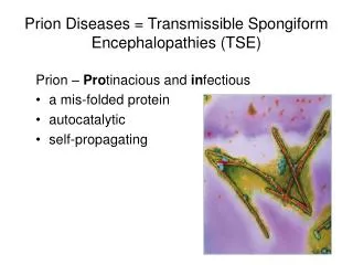
Prion diseases • Infectious neurodegenerative disease with long incubation time • Examples: • Scrapie Sheep • BSE Cattle (Bovine Spongiform Encephalopathy) • TME Mink • CWD Deer, Elk (Chronic Wasting Disease) • FSE Cats • CJD Human (Creutzhfeldt-Jakob Disease) • Transmission by consumption of contaminated animal product & blood transfusion from presymptomatic carrier person • Presymptomatic diagnostics of infected animals and people Prevention of spreading the disease
Two approaches for finding biomarker candidates (1) Proteomic analysis of plasma samples along the time-course throughout the incubation time: BL6-RML, FVB/Ncr-RML (2) Time-course gene expression analysis on whole brain homogenates of all combinations 934 DEGs (Differentially Expressed Genes) CNS-specific & potentially secreted proteins(50) Targeted immunoassay on plasma samples
200 400 600 800 m/z Proteomic Analysis of Serum: LC-MS/MS Tryptic peptides Serum Glycoproteins Prion-infected or Control 4wks 8wks Quantify (MS/MS) Isotope labeling (iTRAQ) 2-Dimensional LC Separation 18wks 2wks 114 115 116 117 SCX m/z H2N-LTEVPALVHK-COOH Identify (MS/MS) Fractionation LC-MS Identification of Biomarker Candidates
Difficulty in Studying Serum Proteins * Top 22 proteins : ~99% of serum proteins 2003, Tirumalai, Mol. Cell. Proteomics
Experimental Procedures • 10 triplicate time-point samples (2 wk, 4 wk,…, 20 wk) replicates pooled to one • Two sets of samples (Infected / Control) • 20 pooled samples were combined to 6 iTRAQ groups. • Each group was treated and SCX-fractionated to 6 fractions • Results from 6 fractions were combined for each group • Proteins identified from all 6 sets of experiments were selected for expression profiling 20 16 18 10 12 8 14 14 2 6 4 8 Infected 20 16 18 10 12 8 14 14 2 6 4 8 Control
Identified plasma proteins • Search parameters for Peptide Prophet results • Singly-tryptic peptides were allowed, or • Limited to doubly-tryptic peptides • To estimate protein abundance changes, principles of Protein Prophet applied to analysis of the multiple sets of experiments (Matlab analysis by D. Hwang) • Abundance changes of peptides from same protein were combined (median) • Abundance changes of same peptides from different time-point groups were combined • Abundance changes of same proteins from Infected or Control sample sets during the whole time course • # of unique proteins identified from all 6 groups: • When singly-tryptic peptides were allowed: 195 • When search was limited to doubly-tryptic peptides: 293 • Total: 357
Biomarker Candidates • Criteria: • Fold change (log2 scale) of proteins from infected versus control increase or decrease constantly (minimal fluctuation) • Abundance of proteins throughout time-course changes constantly for both infected samples and control samples
Proteomic analysis on FVB/Ncr plasma • Proteomic data for FVB/Ncr plasma samples are being analyzed with two search parameters. • Proteins with consistent differential expression in BL6-RML and FVB/Ncr-RML plasma samples will be stronger biomarker candidates.
Immunoassay for brain DEGs on plasma samples • From 50 unique CNS-specific & potentially secreted DEGs, Apod was picked for optimizing the immunoassay platform • Also found as differentially expressed in plasma proteomic data • antibodies against mouse Apod are commercially available • Electrochemiluminescence (ECL)-based immunoassay • 96-well format Multi-array plate • ELISA-type sandwich assay with two antibodies (capture antibody / labeled detection antibody)
Electrode 96-well plate
(3) Sulfo-tag + Detection antibody (4) Light emitted upon electrochemical excitation (1) Capture Antibody (2) Target protein in plasma sample
Dose-dependent linear response for mouse Apod proteins in serum!
Immunoassay for candidate biomarkers • Assay for Apod in real plasma samples • Late time-point samples will be used for testing • If test works, all time-point samples from infected or control mice will be assayed together for expression profiling • With recombinant mouse Apod (S. Qin), absolute quantification is also possible. • Assay for other proteins - Selection criteria • Strong differential expression • Known to be in blood • Two antibodies with different epitopes available • Large difference in late time-point samples (Infected vs. Control)
Acknowledgement • ISB: • Leroy Hood • Inyoul Lee • Daehee Hwang (Now at Postech (S. Korea)) • David Baxter • Nils Gehlenborg • Brianne Ogata (Now at PNNL) • McLaughlin Research Institute: • George Carlson • Doug Spicer • Rose Pitstick