Download
1 / 31
330 likes | 689 Views
Janeway et al. (2005) Immunobiology: The Immune system in health and disease Garland Publishing, 6th edition, figure 3-8. Laboratory uses for antibodies: Immunoprecipitation. Enyzme-linked immunosorbent assay (ELISA) to detect anti-HIV-1 p24 antibodies. Microtitre well coated with p24.

E N D
Janeway et al. (2005) Immunobiology: The Immune system in health and disease Garland Publishing, 6th edition, figure 3-8
Enyzme-linked immunosorbent assay (ELISA) to detect anti-HIV-1 p24 antibodies Microtitre well coated with p24 Serum added Anti-human antibody labeled with enzyme added Enzyme substrate added Note: False positives can result from cross-reactions. See Figure 49.15 in Freeman 3/E
-gp160 -gp120 -p66 -p55 -pg41 -p32 -p24 -p17 Western blot to detect anti-HIV-1 antibodies Lyse (break open) laboratory strain ofHIV-1 in detergent Separate HIV proteins by SDS-PAGE* Transfer proteins to membrane (blot) Cut membrane and incubate in blood sample from individual being tested 6 30 Days after HIV-1 infection Detect bound antibodies SDS is a detergent that binds to and unfolds proteins; PAGE: polyacrylamide gel electrophoresis
Clicker question 1) The polypeptide backbone is negatively charged. 2) The sidechains are negatively charged. 3) SDS is negatively charged. 4) Migration is random; ~half of proteins migrate towards the positive electrode. In SDS-polyacrylamide gel electrophoresis (SDS-PAGE), proteins migrate towards the positive electrode because:
Laboratory uses for antibodies: Immunofluorescence microscopy and flow cytometry Kuby, Kindt, Goldsby, Osborne Immunology Textbook
A- cells A+ cells Single color FACS analysis (e.g., using anti-A antibody) -- note this is a log scale Flow cytometry --Fluorescence Activated Cell Sorting (FACS) A two-color FACS analysis Kuby, Kindt, Goldsby, Osborne Immunology Textbook
Stanford used flow cytometry to screen blood before HIV tests were available Reduced ratio of CD4+ to CD8+ T cells in AIDS patients July 1983 to June 1985, Stanford Blood Center used flow cytometry to test donated units for CD4:CD8 ratio Did not transfuse blood from donors with CD4:CD8 ratio < 0.85 Most other blood banks did no screening ~10,000 cases of transfusion-transmitted AIDS in US before HIV test available in 1985 Galel et al., 1995, “Prevention of AIDS transmission through screening of the blood supply” Annu. Rev. Immunol. 201-227
Another way to sort cells using antibodies Kuby, Kindt, Goldsby, Osborne Immunology Textbook
Having two identical antigen binding sites allows antibodies to bind tightly to surfaces with repeating epitopes Fc Note -- Fc regions of antibodies are exposed when Fabs bind to surface antigens.
IgM IgG Fab IgG Affinity versus avidity Moderate affinity when only one Fab is bound Higher “apparent” affinity due to avidity effects Affinity is a measure of the strength of a binding interaction: A + B <--> AB Equilibrium dissociation constant (KD) = [A][B]/[AB] Strength of binding between a multivalent protein and an antigen or ligand containing multiple binding sites is the AVIDITY. High avidity can compensate for low affinity. Fab IgG No effects of tethering to a surface if only one Fab is bound No avidity effects if antigen is not tethered to a surface
What antibodies do • Don’t directly kill anything • Can block entry of a virus into a host cell or prevent virus from replicating (neutralizing antibodies) gp41 gp120 IgG CD4 CCR5
What antibodies do • Can tag invaders for destruction (first of three ways) • By complement -- binding of IgM or IgG to repeated epitope on invader surface triggers “classical” pathway (movie in extra material at end of Antibodies Lecture 1) Electron micrographs of ~100 Å diameter membrane attack complex channels that are one of the end results of complement activation.
What antibodies do • Can tag invaders for destruction (2nd of three ways) • By macrophages: antibodies opsinize (decorate the surface of) invaders -- Fc receptors on macrophages bind to exposed Fcs to increase phagocytosis
What antibodies do • Can tag invaders for destruction (3rd of three ways) • By Natural Killer (NK) cells: Fc receptors on NK cells bind to exposed Fcs to activate Antibody-Dependent Cellular Cytotoxicity (ADCC) • NK cells bear surface Fc receptors (CD16). If Fc regions of IgG are clustered or aggregated by antigen on a target cell, they bind to CD16. • Binding to CD16 causes contents of granules inside cells to be released --> lysis of target cells.
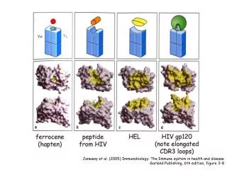
Antibodies can bind to different epitopes on the same antigen -- The immune response to any antigen is polyclonal Protein antigens Note that these three Fabs bind to different regions of the model antigen lysozyme Figure courtesy of Ian Wilson, Scripps Institute Haptens What if you want a single, chemically-homogeneous antibody against an antigen? Answer: you make a monoclonal antibody -- see next slide and link to Köhler and Milstein’s 1984 Nobel Prize lecture on Bi1 website.
Figure A-14 part 1 of 2 B cell hybridomas secrete monoclonal antibodies • Polyclonal B cells secreting antibodies against antigen A cannot be grown in tissue culture, so can’t produce a clone secreting a single type of antibody. • Fuse B cells with myeloma (malignant tumor) cells. These cells have been immortalized (can be grown in tissue culture). • Use myeloma cells that lack the enzyme hypoxanthine:guanine phosphoribosyl transferase (HGPRT) enzyme. • Resulting hybrid cells (hybridomas) secrete antibody and can be grown in tissue culture. • Make clones of single hybridomas.
Monoclonal antibodies have many uses in biology, biotechnology, medicine • Used to detect presence and/or quantity of an antigen; e.g., Western blot, ELISA, immunofluorescence microscopy, immunoelectron microscopy, flow cytometry. • Used to purify antigens; e.g., immunoprecipitation (e.g., CHiP), immunoaffinity chromatograpy. • Used for medical applications, especially for the treatment of cancer. 160 different monoclonal antibodies in clinical trials or awaiting FDA approval (August 2006).
Examples: Therapeutic uses of monoclonal antibodies Rituximab (Genentech) -- against CD20 antigen on surface of normal and malignant B cells. Used to treat non-Hodgkin’s lymphomas (B cell lymphomas). Herceptin (Genentech) -- against HER2 antigen. Given to patients with metastatic breast cancer whose tumors overexpress the HER2 protein (growth factor receptor). (HER2-positive breast cancers are more aggressive than HER2-negative breast cancers.)
Figure 14-17 Different ways monoclonal antibodies are used to eliminate tumors e.g., ricin or Pseudomonas toxin Can also link antibody to a chemotherapy drug (e.g., adriamycin) (ADCC)
Figure A-15 In vitro selection to produce human monoclonal antibodies or increase affinity of existing monoclonal antibody Clone into a phage so that each phage expresses one VH-VL surface fusion protein. Multiply phage display library in bacteria, bind phage to surface coated with antigen. Wash away unbound phage. Repeat procedure (multiply recovered phage, bind to antigen, wash away unbound phage) for several cycles. Recover specific high-affinity antigen binding VH-VL regions. Generate library of heavy and light chain variable regions using spleen DNA. Or introduce random mutations into variable regions genes of a specific antibody.
Clicker question • Epitope : CDR • 1) HV region: Ag • 2) Ab : Ig • 3) Fab : Fc • 4) Ag : Ab
Clicker question After an egg is fertilized, the DNA in the egg is copied. Copies are passed to daughter cells, copied again, passed to new daughter cells, etc. etc. With the exception of errors arising during copying (mutations), all somatic* cells end up with same DNA as the fertilized egg. *somatic = not a germline cell; i.e., not sperm or egg 1) True? 2) False?
Figure 1-18 The diversity of antigen receptors in both B and T cells is generated by rearrangements of gene segments Antibodies (and T cell receptors) are encoded by sets of gene segments. During development of a B (or T) cell, gene segments are joined randomly by DNA recombination (irreversible). Juxtaposed gene segments encode variable part of the antibody (or T cell receptor). Different cells join gene segments differently, so receptors are unique. Each B cell bears many copies of its unique receptor (membrane-bound antibody). Each T cell bears many copies of its unique receptor (T cell receptor; TCR).
V regions encoded by >1 gene segment (light chains) Important point: Rearrangement for antibody genes is at the DNA level -- different from RNA splicing, which occurs in many genes Kuby Immunology textbook
V regions encoded by >1 gene segment (heavy chains) Kuby Immunology textbook
Complement activation - classical pathway Don’t need to memorize the details. Anaphylatoxin: peptides produced during complement fixation that mediate inflammation. Induce anaphylactic shock when injected into animals.
Tetanus – only vaccine-preventable disease that is infectious, but not contagious Tetanus caused by Clostridium tetani, an anaerobic bacterium C. tetani produces endospores, survival structures that begin to metabolize and cause infection once inside an adequate environment Rough surface of a metal is a good place for a C. tetani endospore Stepping on a nail found outdoors (doesn’t have to be rusty) can lead to tetanus if the object causing the puncture wound also delivers endospores Bacteria express a neurotoxin (tetanus toxin or tetanospamin), which is released from dead bacteria inside a host. Activated tetanus toxin carried to spinal cord and brain stem, binds to receptors and blocks neurotransmission Recovery from naturally-acquired tetanus rarely results in immunity because the toxin is extremely potent. Vaccine given as a purified, inactivated toxin (tetanus toxoid). Boosters given every ten years or to anyone with a puncture wound Booster may not work fast enough – can be combined with passive immunization with anti-tetanospasmin antibodies