Download
1 / 1
10 likes | 186 Views
Electroporation Sensitivity of Oxidized Phospholipid Bilayers Zachary A. Levine 1,2 , Yu-Hsuan Wu 3 , Matthew J. Ziegler 1,3 , D. Peter Tieleman 4 , and P. Thomas Vernier 1,5.

E N D
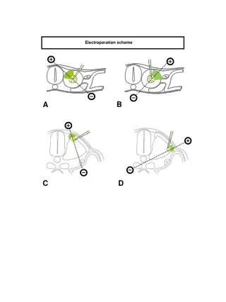
Electroporation Sensitivity of Oxidized Phospholipid Bilayers Zachary A. Levine1,2, Yu-Hsuan Wu3, Matthew J. Ziegler1,3, D. Peter Tieleman4, and P. Thomas Vernier1,5 1 MOSIS, Information Sciences Institute, Viterbi School of Engineering (VSoE), University of Southern California (USC), Marina del Rey, USA 2 Department of Physics and Astronomy, College of Letters, Arts, and Sciences, USC, Los Angeles, USA 3 Mork Family Department of Chemical Engineering and Materials Science, VSoE, USC, Los Angeles, USA 4 Department of Biological Sciences, University of Calgary, Calgary, Canada 5 Ming Hsieh Department of Electrical Engineering, VSoE, USC, Los Angeles, USA, Molecular Dynamics Results 2 Introduction Electroporation time for 50% oxidized systems versus applied electric field Top View of a Quilted PLPC Bilayer Containing Localized oxPLPC Clusters Molecular dynamics (MD) studies showing that oxidized lipids increase the frequency of water defects in phospholipid bilayers suggest that the presence of oxidized lipids in a bilayer will also increase the sensitivity of the bilayer to electropermeabilization. We confirmed this hypothesis in MD simulations of PLPC (1-palmitoyl-2-linoleoyl-sn-glycero-3-phosphatidylcholine) bilayers containing oxidized PLPC (oxPLPC), showing that pore creation is affected by the oxPLPC species, its concentration in the bilayer, and the electric field strength. We also demonstrated that these effects can be localized to a specific region of the bilayer. Experimental observations with living cells are consistent with the simulations. Methods 13-tc oxPLPC PLPC 12-al oxPLPC All simulations were performed using the GROMACS 3.3.1 package and all initial oxPLPC systems were derived from previous work on oxidized lipids [1]. The systems were composed of PLPC plus oxPLPC lipids (with 12-oxo-cis-9-dodecenoate (12-al) or 13-hydroperoxy-trans-11,cis-9-octadecadienoate (13-tc) at the sn-2 position) in concentrations of 0%, 11%, 25%, or 50% of the total system, with the oxPLPC lipids distributed equally in the two leaflets of the bilayer. Each system contained 72 lipids and 2880 water molecules (40 waters per lipid) and was energy minimized and equilibrated for 80 ns. A simulation with a larger area was created by doubling a 72-PLPC bilayer in x and y and then individually replacing PLPC lipids with oxPLPC lipids in two opposing quadrants, creating a quilted system where two quadrants are heavily oxidized (50% oxPLPC) and the two remaining quadrants contain only PLPC. This enables us to test whether electroporation occurs preferentially in oxidized regions of a bilayer. Periodic boundary conditions were employed to mitigate system size effects and the integration time step was 2 fs. Short-range electrostatics and Lennard-Jones interactions were cut off at 1.0 nm. Long-range electrostatics were calculated by the particle mesh Ewald (PME) algorithm using fast Fourier transforms and conductive boundary conditions. Electroporation times were calculated by identifying the first simulation step in which any phosphorus atom in one leaflet approached within 1.2 nm of any phosphorus atom in the other leaflet. Blue – Nitrogen Gold – Phosphorus Red – Oxygen Cyan – Carbon (Single Bond) Black – Carbon (Double Bond) The time to create a hydrophilic pore decreases as the electric field increases, with 12-al oxPLPC more susceptible to electroporation than 13-tc. Each simulation ran for a total of 25 ns. Before an electric field is applied After an electric field is applied Shown here is our large quilted system which contains PLPC (white) and selectively placed oxPLPC (blue). The system is a 4x4 array of sectors containing alternately pure PLPC and 50% oxPLPC (12-al). After an electric field is applied, there is a clear correlation between pore location and local oxidation clusters. Molecular Dynamics Results 1 Electroporation time versus oxidized lipid concentration Live Cell Results Composite oxPLPC snapshots taken at 0.5 ns intervals over a total time of 10 ns. Facilitation of water entry into the bilayer interior by oxPLPC aldehyde or hydroperoxy oxygens Pulse-Induced YO-PRO-1 Uptake in Peroxidized Jurkat Cells (30 ns, 3 MV/m, 50 Hz) all lipids shown oxPLPC sn-2 tails shown 12-al oxPLPC Mean Phosphorus Plane oxPLPC lipids have a tendency to bend their sn-2 tails towards the aqueous interface. This process does not appear to be affected by the presence of an external electric field, though 13-tc oxPLPC appears to bend much more towards the aqueous interface than 12-al oxPLPC. Normal, 0 pulses Normal, 50 pulses 1.3 ns 1.3 ns Peroxidized, 0 pulses Peroxidized, 50 pulses Pulse-induced YO-PRO-1 uptake in peroxidized cells is significantly higher than in control cells (1.8X in cells treated with 500 µM H2O2, 1 mM Fe2+, then 50 pulses). 13-tc oxPLPC Cells were treated with peroxidizing reagents for 10 minutes and pulsed immediately without washing. Pore creation time decreases as the concentration of oxidized lipids increases. A field of 0.36 V/nm was used in all systems because it corresponded to a ‘minimum porating field’ for PLPC, a field we have previously defined as one which electroporates a system in one of three trials within 25 ns [2]. Mean Phosphorus Plane 10.6 ns 14.6 ns Conclusions Simulated bilayers containing oxidized lipids have an increased susceptibility to electroporation. This increase in susceptibility is likely due to the facilitation of water transport into the bilayer interior by hydroperoxy or aldehyde oxygens on the oxidized residues and may also be a consequence of the fact that oxPLPC bilayers are thinner than pure PLPC bilayers. The presence of an electric field does not appear to drastically change the average distance ofaldehyde or hydroperoxy oxygens to the aqueous interface. Clusters of oxPLPC lipids attract a large number of individual waters into the bilayer interior,creating localized regions of electroporation susceptibility. MD results are consistent with experimental observations. Cells treated with peroxidizing agentsappear to electroporate more readily than untreated cells. Average distance (nm) from tail oxygen or PLPC C13 to the mean phosphorus plane No Field Field* [1] J. Wong-ekkabut, et al. Effect of Lipid Peroxidation on the Properties of Lipid Bilayers: A Molecular Dynamics Study. Biophys. J., 93: 4225–4236, 2007. 13-tc 0.34 0.32 [2] M.J. Ziegler and P.T. Vernier. Interface Water Dynamics and Porating Electric Fields for Phospholipid Bilayers. J. Phys. Chem. B, 112(43), 13588–13596, 2008. 12-al 0.60 0.73 1.54 1.59 PLPC Special thanks to the USC Center for High Performance Computing and Communications (HPCC) for providing the computational resources, and to MOSIS for providing funding. 16.6 ns 16.9 ns *At a strength of 0.36 V/nm Successive snapshots of electropore formation in an 11% 12-al oxPLPC system. Average water penetration depth corresponds to the average depth of oxygen atoms on the sn-2 tail. Only oxidized sn-2 tails and water are shown.