Download
1 / 20
200 likes | 429 Views
Chpt 14. The digestive system & metabolism. Important terms. Ingestion – taking in of food Digestion – breaking down of food, both physically & chemically Absorption – movement of nutrients throughout the bloodstream Defecation – ridding the body of indigestible wastes

E N D
Chpt 14 The digestive system & metabolism
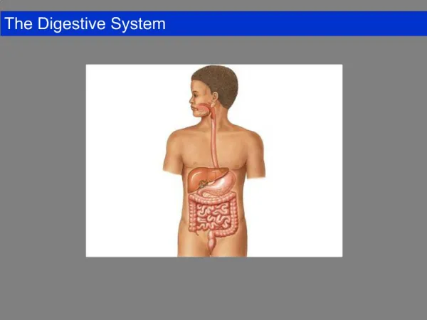
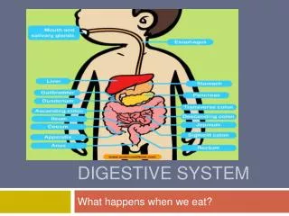
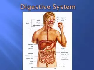
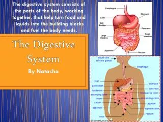
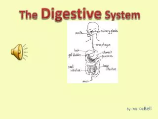
Important terms • Ingestion – taking in of food • Digestion – breaking down of food, both physically & chemically • Absorption – movement of nutrients throughout the bloodstream • Defecation – ridding the body of indigestible wastes • Alimentary canal – true “digestive tract” – the path food takes once it enters the mouth and exits the anus • Accessory organs – help with the process of digestion, but food does not actually travel there • Teeth, salivary glands, pancreas, liver, gallbladder
Organs of the Alimentary Canal: Mouth • Lips (labia)—protect the anterior opening • Cheeks—form the lateral walls • Hard palate—forms the anterior roof • Soft palate—forms the posterior roof • Uvula—fleshy projection of the soft palate • Vestibule—space between lips externally and teeth and gums internally • Oral cavity proper—area contained by the teeth • Tongue—attached at hyoid bone and styloid processes of the skull, and by the lingual frenulum to the floor of the mouth
Physiology of the Mouth • Mastication = chewing • Saliva is released once food is masticated • Initiation of swallowing by the tongue • Allows for sense of taste
Esophagus • About 10 inches long (3 lengthwise fists) • Runs from the pharynx to the stomach, through the diaphragm • Moves food to the stomach through rhythmic contractions called peristalsis • Passageway for food only • Respiratory system branches off after the pharnyx (hence the larnyx only serves a respiratory function)
Ogars have layers, onions have layers • All alimentary canal organs have many layers • Mucosa • Innermost , moist membrane • Surface epithelium • Small amount of connective tissue & smooth muscle • Submucosa • Just beneath the mucosa layer • Soft connective tissue containing • Blood vessels, nerve endings & lymphatic tissue • Muscularisexterna • Two layers of smooth muscle • Serosa • Outermost wall containing fluid producing cells • Parietal peritoneum: lines the abdominopelvic cavity • Visceral peritoneum: outermost layer that is connected to parietal peritoneum
Stomach • Located on the left side of the abdominal cavity • Food enters at the cardioesophageal sphincter • Food empties into the small intestine at the pyloric sphincter (valve) • Regions of the stomach • Cardiac region—near the heart • Fundus—expanded portion lateral to the cardiac region • Body—midportion • Pylorus—funnel-shaped terminal end • Rugae—internal folds of the mucosa
Stomach Physiology & Mucosa Structure • Temporary storage tank for food • Site of food breakdown • Chemical breakdown of protein begins • Delivers chyme (processed food) to the small intestine • Mucosa is simple columnar epithelium • Mucous neck cells—produce a sticky alkaline mucus • Gastric glands—situated in gastric pits and secrete gastric juice • Chief cells—produce protein-digesting enzymes (pepsinogens) • Parietal cells—produce hydrochloric acid • Enteroendocrine cells—produce gastrin
Hormones that act in digestion • All the hormones below are made by the stomach mucosa & stimulated by food entering the stomach. • Gastrin: • Stimulates the emptying of the stomach, stimulates contraction of intestinal muscles • Relaxes ileocecal valve • Stimulates mass movement of the contents of the large intestine • Serotonin: • Causes contractions of the stomach muscle • Histamine: • Activates the parietal cells to release HCl (starting the digestion process) • Somatostatin • Inhibits secretion of all gastric products (important for negative feedback) • Slows down blood flow to intestines & decreases intestinal absorption • Inhibits the release of bile from the gallbladder
Small Intestine & its subdivisions • The body’s major digestive organ • Site of nutrient absorption into the blood • Muscular tube extending from the pyloric sphincter to the ileocecal valve • Suspended from the posterior abdominal wall by the mesentery • Duodenum • Attached to the stomach • Curves around the head of the pancreas • Jejunum • Attaches anteriorly to the duodenum • Ileum • Extends from jejunum to large intestine
Chemical Digestion in the Small Intestine • Chemical digestion begins in the small intestine • Enzymes are produced by • Intestinal cells • Pancreas • Pancreatic ducts carry enzymes to the small intestine • Bile, formed by the liver, enters via the bile duct
Small Intestine Anatomy • Three structural modifications that increase surface area • Microvilli—tiny projections of the plasma membrane (create a brush border appearance) • Villi—fingerlike structures formed by the mucosa • Circular folds (plicaecirculares)—deep folds of mucosa and submucosa
Hormones that act in digestion • All hormones listed below are produced by the duodenal mucosa • Intestinal gastrin: stimulated by acidic & partially digested foods entering the duodenum • Stimulates secretion of gastric products & intestinal motility • Secretin: stimulated by acidic chyme • Inhibits secretion of gastric products • Increases output of pancreatic juices to breakdown carbohydrates and proteins • Increases bile output • Cholecystokinin (CCK): stimulated by fatty chyme • Aids in the effectiveness of bile • Increases output of pancreatic juice • Stimulates the gallbladder to expel bile • Relaxes the hepatopancreatic sphincter to allow entry of bile into duodenum
Hormones that act in digestion • Gastric inhibitory peptide (GIP): stimulated by fatty or glucose-containing chyme • Inhibits gastric gland secretion & motility • Vasoactive inhibitory peptide (VIP): chyme containing all kinds of partially digested foods • Stimulates buffer secretion • Dilates intestinal capillaries to prepare for absorption of nutrients • Inhibits HCl production (stops further digestion) • Relaxes the intestinal smooth muscle to increase surface area for absorption
Large Intestine • Larger in diameter, but shorter in length, than the small intestine • Frames the internal abdomen • Cecum—saclike first part of the large intestine • Appendix • Accumulation of lymphatic tissue that sometimes becomes inflamed (appendicitis) • Hangs from the cecum
Large Intestine • Colon • Ascending—travels up right side of abdomen • Transverse—travels across the abdominal cavity • Descending—travels down the left side • Sigmoid—enters the pelvis • Rectum and anal canal—also in pelvis • Anus—opening of the large intestine • External anal sphincter—formed by skeletal muscle and under voluntary control • Internal involuntary sphincter—formed by smooth muscle • These sphincters are normally closed except during defecation
Large Intestine • No villi present • Goblet cells produce alkaline mucus which lubricates the passage of feces • Muscularisexterna layer is reduced to three bands of muscle called teniae coli • These bands cause the wall to pucker into haustra (pocketlike sacs)
Liver & gallbladder • Work together to create & store bile • The liver ejects bile directly into the duodenum to emulsify fats • When fats are broken down into tiny particles, they are more easily digested • Liver – appx. 3 lbs • Extends from the right hypochondriac to epigastric regions • Divided into 4 lobes with its own specific circulation (hepatic portal) • Entire liver is covered by serosa (visceral peritoneum), which attaches the liver to the diaphragm • Gallbladder is chiefly responsible for storage of bile
Gallbladder • Thin walled, green, muscular sac • About the size of a kiwi (4 inches) • Gland that extends from the interior margin of the liver • Stores bile that is not needed immediately for digestion • Since it is not needed immediately, the gallbladder concentrates that bile by absorbing some of its water content • When empty or only containing a small amount of bile, it can fold up like the rugae of the stomach to allow for quick expansion and expulsion of bile when needed
Pancreas • Extends across the abdomen • Lies deep to the greater curvature of the stomach • Produces a broad spectrum of enzymes to breakdown foodstuffs • Pancreas delivers pancreatic juice directly to the duodenum • Usually fuses with the bile duct from the liver/gallbladder • Pancreatic juice mainly consists of water, electrolytes, and enzymes • Has a high pH – enables neutralization of acidic chyme • Pancreatic enzymes include: • Amylase, lipases, nucleases • Carboxypeptidase: broad range of functions: mainly protein digestion • Chymotrypsin: breaks down proteins: specifically: tyrosine (cheese protein), tryptophan & phenylalanine • Enterokinase – a serine protease – breaks bonds between peptides