Download
1 / 8
90 likes | 221 Views
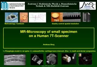
Zentrum f. Medizinische Physik u. Biomedizinische Technik & MR- Hochfeld - Zentrum. MR- Microscopy of small specimen on a Human 7T-Scanner Andreas Berg. Methodology : hardware. Quality control spatial resolution.

E N D
Zentrum f. MedizinischePhysik u. BiomedizinischeTechnik & MR-Hochfeld-Zentrum MR-Microscopyofsmallspecimenon a Human 7T-ScannerAndreas Berg Methodology: hardware Quality control spatial resolution 1.) Paraplegia model in rat spine 2.) osteoarthritis: cartilage bone intervert. Disc 3.) teeth and dental composites A.Berg, MR-Center 06.04.2011
ResultsQuality control: Spatial Resolution Standard whole body scanner: Phantoms: Test structures d > 1000 µm MR-microscopyinsert on 7T scanner: 1,5 cm A. Berg, Deutsches Patentamt, München, DE19904635 , 19.08.1999 Phantoms: teststructures d < 100 µm
First results: Quality Control: spatial Resolution Silicon grid in Silicon oil TM = 87 min SNR = 16 Voxelsize: 50,9 x 25,4 x 200 µm3 !!! = 2,6 10-4 mm3 = 0,26 10-9 l < 10-3 Clinical Voxel size (500 x 500 x 2000 µm) TM = 1,5 min SNR = 3,15 MR-microscopic images of Silicon grid sets in Silicon oil (finest: a/2 = 32 µm) Left: sagittal view at increased SNR (32 averages ) Top: enlarged area trace of profile is indicated Bottom: profile indicating modulation depth Right: sagittal view for a short protocol A. Berg et al. MR-MICROSCOPY ON A HUMAN 7T-SCANNER; ISMRM/ESMRMB 2010, progrnr. 1048,
MR-microscopy vs. histology: Osteoarthritis MR-microscopy: A. Berg; Samples andreferencing: S. Domayer, J. Hofstätter Univ. klinik f. Orthopädie
Osteoarthritis cartilage-boneinterface: MR-microscopyvshistologicalstaining 1.) Healthycartilage(lateral condyleoftheknee): Homogeneous, thickcartilagelayer • MR-microscopyvshistology: • Intacttissue after measurement • Nodamage due tocutting • Nostainingnecessary • (T2, T1, Diffusivity) • Low effort, • Low preparation - andmeasurement time trabecularnetworkstructure MR-microscopy: TE = 8 ms Voxel: 96 x 59 x 240 µm3 A. Berg, A. Potthast, P. Starewicz, S. Domayer, J. G. Hofstaetter; MR-Microscopy on a human MR-7T Scanner: Methodologyandapplicationto medial osteoarthriticsamplesoftheKnee. ECR 2011 Vienna Histologicstaining: Safranin-O
MR-Microscopyvs. histology osteoarthriticcartilage(medial condyleoftheknee): 8 mm 5 MR-microscopy: Voxelsize = 51 x 51 x 240 µm3 < 10-3clinical MRI voxelsize (500 x 500 x 2000 µm3 ) TSE: 3D-MTX: 256x 256 x 47 TE = 8.2 ms, TR = 2.3 s TM = 16 min Size: 250 µm - 50 µm Small degradationareasbelowthesurface Presumably: areasoffibrillationandlossofproteoglycane A. Berg, A. Potthast, P. Starewicz, S. Domayer, J. G. Hofstaetter; MR-Microscopy on a human MR-7T Scanner: Methodologyandapplicationto medial osteoarthriticsamplesoftheKnee. ECR 2011 Vienna
MR-MicroscopyvsHistology osteoarthriticcartilage(medial compartmentoftheknee): 4 mm Small degradationareasbelowthesurface Presumably: areasoffibrillationandlossofproteoglycane A. Berg, A. Potthast, P. Starewicz, S. Domayer, J. G. Hofstaetter; MR-Microscopy on a human MR-7T Scanner: Methodologyandapplicationto medial osteoarthriticsamplesoftheKnee. ECR 2011 Vienna
3D-multislicing 13 mm Pulse sequence: Berg_TSE Voxel: 51 x 51 x 240 µm3 3D-MTX: 256x 256 x 47 TM = 16 min A. Berg, A. Potthast, P. Starewicz, S. Domayer, J. G. Hofstaetter; MR-Microscopy on a human MR-7T Scanner: Methodologyandapplicationto medial osteoarthriticsamplesoftheKnee. ECR 2011 Vienna