Microscopy and Specimen Preparation
630 likes | 1.57k Views
Microscopy and Specimen Preparation. BIO3124 Lecture #2 . Objectives and reading. Reading: Ch.2 1 . Principles of optical resolution of microorganisms Optical behaviour of light Detection, magnification and resolution How to increase resolution Optical parameters Increasing contrast

Microscopy and Specimen Preparation
E N D
Presentation Transcript
Microscopy and Specimen Preparation BIO3124 Lecture #2
Objectives and reading • Reading: Ch.21. Principles of optical resolution of microorganisms • Optical behaviour of light • Detection, magnification and resolution • How to increase resolution • Optical parameters • Increasing contrast 2. Microscopic systems and their applications • Bright field • Dark field • Phase contrast • Fluorescence and Confocal • EM • Scanning Probe (AFM, STM)
Optical behavior of light • Interaction of light with objects • Absorption: object will appear dark • Reflection • Refraction: basis for magnification • Scattering: when size of object is close to the wavelength of light • Principle: to be resolved the wavelength should be smaller than the size of object
Detection, Magnification and Resolution Definitions • Optical systems: detect, magnify, resolve • Eye, simple lens, microscopes • Use electromagnetic radiation, eg. visible light, laser or electron beam • Detection:ability to determine the existence of an object • Resolution: ability of an optical system to distinguish two small close objects • Human eyes limit of resolution is 150 µm • Magnification: ability to increase the apparent size of the image of an object
10 mm Observing Microbes • Microscope needed to see smaller objects • Eukaryotic microbes • Protozoa, algae, fungi • 10–100 mm • Prokaryotes • Bacteria, Archaea • 0.2–10 mm • Viruses • 0.01-0.1 mm Lactobacillus lactis Amoeba proteus Poliovirus
Magnification • Microscopes: Magnify image to match the limit of resolution of eye retina ie. 150 um • Magnification’s contribution to resolution is limited • Distortion due to light wave interference • Empty magnification: magnification that does not improve resolution • Magnification is due to refraction • Refraction: light is refracted (bent) at the varying density interfaces • Light travels slower, waves compressed (higher frequency) • Depends on refractive index of object
Lenses and the bending of light • Refractive index (RI) • how greatly a substance slows the velocity of light • Direction/magnitude of bending depends on the RI • A lens behaves like a prism Normal Incident angle Refracted angle
Refraction and Magnification • Image forms at crossing refracted light originated from the object • Magnification ratio depends on the position of object with respect to the lens Watch tutorial
Light Microscopes • Compound microscopes • image formed by action of 2 lenses • Bright-field microscope • Dark-field microscope • Phase-contrast microscope • Fluorescence microscope
The Bright-Field Microscope • Dark image against a brighter bkg • Several objective lenses Parfocal: stays focused when objectives changed • total magnification (max 1000-fold) • product of the magnifications of the ocularrlensesand the objective lenses
Microscope Resolution • Optical parameters affecting resolution • Shortest distance resolved by an optic system (d) is expressed by: • Abbe equation: d=0.5λ/n.sinθ • λ= wavelength, n= refractive index, θ= angle of apreture • shorter wavelength greater resolution • Numerical aperture: NA= n.sinθ • Smaller d value = more powerful optic system
Microscope Resolution Reducing the d value (higher resolution) means increasing the θ, • working distance • distance between the front surface of lens and surface of coverslip or specimen when it is in sharp focus
Microscope Resolution • Effect of refractive index: NA= n.sinθ
The Dark-Field Microscope • Image is formed by light reflected or refracted by specimen • Interference by the bkg light eliminated • produces a bright image against a dark bkg • to observe living, unstained preparations • For eucaryotes has been used to observe internal structures • For procaryotes has been used to identify bacteria such as Treponema pallidum, the causative agent of syphilis
Dark field microscopy: Light path • Spider Light stop: produce annular ring of light, no light from the centre enters objective • only light passing through object enters the objective lens • bkg stays dark, specimen shines
Dark field microscopy Example of an insect larva examined in a dark field microscope
Frits Zernike (1888-1966) The Phase-Contrast Microscope • first described in 1934 by Dutch physicist Frits Zernike • enhances the contrast btw intracellular structures that have slight differences in their refractive indices • excellent tool to observe living cells • bacterial components such as endospores and inclusion bodies • Eukaryotic organelles
HeLa cells Reza Nokhbeh Phase contrast image of HeLa cells
Phase contrast microscopy P.aeruginosa Sporulating bacterium Contrast between spores and Vegetative forms Paramecium Intracellular organelles contrasted
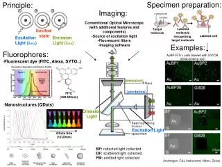
The Fluorescence Microscopy • specimens usually stained with antibodies tagged with a fluorophore • Excitation light: ultraviolet, violet, or blue light activates fluorophore tagged cells • Emission light: longer wavelength, enters objective • bright image of the object resulting from the fluorescent light emitted by the specimen • Applications: medical microbiology and molecular biology
Infected Poliovirus infected HeLa T4 cells Reza Nokhbeh Poliovirus interferes with the integrity of SiRNA centres • GW bodies disintegrate as the result of Poliovirus infection • virus and GW bodies are stained with fluorochrome conjugated specific antibodies
Electron Microscopy • Ernst Ruska and Max Hall in Germany finished the first prototype in 1931 • Eli Franklin Burton (1847-1948) and his students, James Hillier, Cecil Hall and Albert Prebus, built the first functional EM in 1938 at Toronto university • Louis de Broglie’s principle that electron particles also have electromagnetic (wave) property • accelerated electronic beam in microscopy would enhance resolution, why? James Hillier (1915-2007)
Transmission Electron Microscopy (TEM) • wavelength of electron beam is much shorter (0.005 nm or 5 A˚) than light, i.e. much higher resolution • Magnification is 100,000 to 200,000 • Resolution approaches 0.5 nm, ie about 1000-fold higher than light microscopes
Principles of light microscopy applies to TEM Thermionic Electron Gun ~300 Kev monochromatic beam
The Scanning Electron Microscopy (SEM) • uses electrons scattered from the surface of a specimen to create image • produces a 3-dimensional image of specimen’s surface features
Examples of TEM and SEM micrographs P. acens lytic phage TEM, 150,000x R. Nokhbeh , J. Trifkovic
New Techniques in Microscopy • Confocal laser scanning microscopy(CLSM) and scanning probe microscopy • have extremely high resolution • Expanded the resolution to molecular and atomic levelsi.e. 1-100 A
Confocal Microscopy Confocal Laser Scanning Microscope (CLSM) • laser beam used to illuminate a variety of planes in the specimen, exciting fluorophore • computer compiles images to generate 3D image • used extensively to observe biofilms • Also used in studying the sub-cellular structures • Light is only gathered from the plane of focus
Confocal scanning laser microscope • blurring does not happen since signal is gathered by scanning a thin • layer of specimen, plane of focus, at each round
Scanning Probe Microscopy • Atomic Force Microscope (AFM) • Vertical movement of probe is followed by a laser beam • probes surfaces that are not charged
Atomic Force Microscope Membrane integral aquaporin protein captured by AFM α-synuclein protein fibers. Misfolded fibers are incolved in Parkinson disease Human mitotic chromosome spread
Scanning Probe Microscopy • Scanning Tunneling Microscope (STM) • Measures the surface features of specimen by moving a sharp probe over the surface • steady current (tunneling current) maintained between microscope probe and specimen • up and down movement of probe as it maintains current is detected and used to create image of surface of specimen • Magnification: 100 million times, capable of detecting the surface atoms
Atoms of MoS2, the bright spots are S atoms DNA double helix Scanning Tunneling Microscope Silicon surface atoms enlarged 20 million times individual surface atoms and the bonds that hold them in place
Preparation and Staining of Specimens • Staining techniques are applied to increase the contrast • increases visibility using bright field microscopes • accentuates specific morphological features • preserves specimen (due to fixation)
Fixation • preserves internal and external structures and stabilizes them in position • organisms usually killed and firmly attached to microscope slide • heat fixation – routinely used in procaryotes, preserves overall morphology but not internal structures • chemical fixation – used for larger, more delicate organisms • protects fine cellular substructure and morphology
Dyes • Dyes • Ionizable dyes have charged groups Cationic (basic):Positively charged. • e.g. Methylene blue, Crystal violet, Safranine, Malachite green. Anionic(acidic):Negatively charged • e.g. Nigrosin black, Indigo ink.
Simple and Differential staining • Simple staining • a single stain is used • use can determine size, shape and arrangement of bacteria • Differential staining divides microorganisms into groups based on their staining properties • e.g., Gram staining • e.g., acid-fast staining
Staining • Positive staining: Specimen staining.
Staining (Contd) • Negative staining: • Background staining, not the specimen.
Methods Simple Staining: • One type of stain. • Cationic or Anionicstains. • Able to determine the size, shape and the arrangment of bacteria.
Different Cell Morphologies • Coccus: • Sphere • 3 planes of division • Plane of division producesdifferent arrangements of cells. • Typical arrangements for differentbacterial types. • Bacillus: • Rods • One plane of division
Cocci Division axes Diplococcus Streptococcus (4-20) Tetrad Staphylococcus
Bacilli (Bacillus) Diplobacilli Streptobacilli
Other Cellular Forms Curved rods (coccobacillus) Vibrio cholerae Spirals Spirochetes
Differential Staining Techniques: Gram Staining • Bacteriadividedintotwo groups: • Gram Negatives: stainred • Bacilli:Escherichia, Salmonella, Proteus, etc. • Cocci:Neisseria and Pneumococcus. • Gram Positives: stainblue/purple • Bacilli:Bacteriafrom the genera of Bacillus and Clostridium • Coccus:Streptococcus, Staphylococcus, Micrococcus