Download
1 / 13
130 likes | 136 Views
European Journal of Experimental Biology is an open access journal, authors and readers reflect a broad interdisciplinary group of scientists who study molecular, cellular and organismal physiology in an evolutionary and environmental context.<br> <br>European Journal of Experimental Biology serves an intellectual audience that is interested in the latest research coming out of universities and industries around the world. The journal publishes research developments and disseminates them to an international audience. The journal welcomes publications of high quality papers. Original research papers, reviews and high quality technical notes.<br><br>Submit manuscript online by registering at Alternatively authors can send articles as attachment to https://www.imedpub.com/submissions/european-experimental-biology.html or send as an e-mail attachment to the editorial office at publisher@imedpub.com<br>

E N D
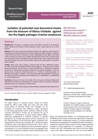
Research Paper 2020 iMedPub Journals www.imedpub.com European Journal of Experimental Biology Vol. 10 No. 6:117 ISSN 2248-9215 Elie Khoury1, Antoine abou fayad2,3,4, Dolla Karam Sarkis5, Mireille kallassy awad1* Isolation of potential new biocontrol strains from the blossom of Malus trilobata against the fire blight pathogen Erwinia amylovora 1 Biotechnology Laboratory, UR EGP, Saint- Joseph University, B.P. 11-514 Riad El Solh Beirut1107 2050, Lebanon Abstract Background: Fire blight is a contagious disease that affects members of the Rosaceae family, caused by the bacteria Erwinia amylovora, which invades apple trees via their blossom. 2 Department of Experimental Pathology, Immunology and Microbiology, American University of Beirut, Beirut, Lebanon Methods: In this study, using culture dependent methods, we isolated for the first time the microorganisms colonizing the blossom of the wild apple tree, Malus trilobata, in Lebanon. A total of 94 strains from the blossoms of trees originating from two regions, Ain Zhalta reserve and Dhour EL Choueir, were isolated. Genetic analysis of the strains revealed a wide and interesting variety of microorganisms quite different from those isolated from Malus domestica blossom. Direct and indirect inhibition assays of the isolates against E. amylovora were conducted. 3 Center for Infectious Diseases Research (CIDR), American University of Beirut. 4 WHO Collaborating Center for Reference and Research on Bacterial Pathogens, American University of Beirut Findings: While some strains belong to species known for their antibacterial activity, two new strains with interesting inhibitory activity against E. amylovora, have not been previously described. The first strain is a fungi, Saccothecium sp., inhibiting E. amylovora growth due to antimicrobial metabolite production and nutrient competition. As for the second strain, a bacterium, Mycolicibacterium sp., it inhibits E. amylovora growth via diffusible antimicrobial. In addition, novel strains potentially competing with E. amylovora for nicotinic acid and nicotinamide were identified such as Filobasidium sp., Rhodotorula sp. and Acinetobacter sp. 5 Laboratory of Pathogens, School of Pharmacy, Saint-Joseph Beirut, Lebanone University, Conclusion: We report here two new isolates from the blossom of Malus trilobata with high potential of biocontrol activity against E. amylovora either via secondary metabolites production and/or by competition at nutrient levels *Corresponding author: Mireille Kallassy Awad Keywords:Fire blight, Erwinia amylovora, Malus trilobata, antibacterial, Mycolicibacterium, Saccothecium mireille.kallassy@usj.edu.lb Faculty of Science, Biotechnology Laboratory, UR EGP, Saint- Joseph University, B.P. 11-514 Riad El Solh Beirut1107 2050, Lebanon Received: July 15, 2020; Accepted: July 22, 2020; Published: August 15, 2020 Introduction Fire blight disease of rosaceous plants, caused by Erwinia amylovora, affects mainly apple, pear trees and many other woody plants of Rosaceae family. This pathogen can be controlled via treatment with antibiotics, copper derivatives or with taking appropriate culture measures. However, certain countries, particularly in Europe, do not authorize the use of antibiotics i.e. streptomycin, to avoid several adverse effects, particularly development of antibiotic resistance in bacterial pathogens [1–3]. An alternative way to control E. amylovora is via the characterization of new biocontrol agents, preferably ones originating from the blossom ecosystem [4]. E. amylovora begins its infection cycle in the stigmas of blossom then, due to rain, it will be transferred to the hypanthium part and invade through the nectarthodes [2, 5]. At the hypanthium level, the pathogen finds the nutrients needed for its growth, which include Nicotinic Acid Citation: Khoury E, Fayad A, Sarkis DK, Awad MK, et al. (2020) Isolation of potential new biocontrol strains from the blossom of Malus trilobata against the fire blight pathogen Erwinia amylovora. Eur Exp Biol Vol.10 No. 6:117 (NiAc) and Nicotinamide (NiNH2). The 6-Hydroxynicotinic acid (6-HNiAc) has been shown as well to be required by E. amylovora as a growth factor at laboratory scale [6, 7]. A study showed that Pseudomonasrhizosphaerae can degrade both NiAc and NiNH2 in a laboratory prepared medium and can colonize apples hypanthia leading to a strong suppression of E. amylovora [7]. Moreover, nicotinic acid and nicotinamide were found two times higher in pear than apples’ hypanthium [6]. Also, the natural defense 1 © Under License of Creative Commons Attribution 3.0 License | This article is available in:http://www.imedpub.com/european-journal-of-experimentalbiology/
2020 European Journal of Experimental Biology Vol. 10 No. 6:117 ISSN 2248-9215 against fire blight attacking the tree through the flower can be due to the flora that resides there competing for nutrients, for example the concentration of sugar on bean leaves decrease to the tenth upon colonization by Pseudomonasfluorescens A506 [8]. Since the phyllosphere is a reservoir for bacteria, fungi and yeasts, several studies focused on the leaves, the dominant surface of the epiphyte [9]. Epiphytic surfaces, as well as blossom stigmas and hypanthium, are considered to be a rich reservoir of diverse microorganisms [10, 11], potential biocontrol agents to fight against E. amylovora [2]. Although several ways to apply isolated biocontrol strains could be adopted, treatment with the antagonistic strain itself showed to be more efficient than its metabolites, since the latter remain on the surfaces of the treated crops whereas the strain will be spread with insects and other factors [5]. Moreover because of the lack of nutrient at epiphytic level, the colonizing strains compete between each other [4]. Therefore, from a biocontrol perspective, not only is the production of metabolites interesting but also the competition for space and nutrients. Hence the importance of searching for new biocontrol agents active against known pathogens such as Erwinia amylovora. Microbial agents can be distinguished by different modes of action, behavior, stability, colonizing capacity and consequently better antagonistic activity. For example, Pseudomonas graminis isolated from Malus spp phyllosphere was highly active against E. amylovora compared to different commercialized antibacterial agents such as: Pantoea vagans C9-1, the bioproducts BlightBan A506 (Pseudomonas fluorescens A506), the blossom protect (Aureobasidium pullulans based) and Hortocyna 18 SP (streptomycin). Pseudomonas graminis colonizes apple blossoms under different weather conditions and throughout the entire blooming period [12]. Other effective yeast biocontrol agents were isolated from Malus pumila blossoms such as Cryptococcus magnus and Pichia guilliermondii [2]. As E. amylovora is a serious plant pathogen for apple trees at the pre- harvest level, the aim of our study was to isolate the culturable microorganisms associated to blossom of the wild endemic apple tree, Malus trilobata (M. trilobata) and testing in vitro their ability to inhibit E. amylovora on plate, directly by metabolite productions or indirectly via nutrient or space competition in order to select new promising biocontrol agents against fire blight disease. These parameters are indicators of blossom maturity age of (4-5 days) where microorganisms are present in large quantity [13]. Collected blossoms were kept inside a sterile plastic container, transported on ice and analyzed 24 hours after sampling. Isolation of microorganisms from the blossom To isolate microorganisms from the blossom, we followed the protocol as previously described by Lawrence et al. 2009 [2] with some modifications. At the laboratory, the blossoms were placed inside a plastic container on ice. The petals were removed using sterile gloves and sterile forceps. Hereafter, every 20 flowers were assembled for dissection. The stigmas of each flower were cut along with a portion of its supporting style and placed in 1 mL phosphate buffer 10 mM, pH 7.0. Dissected stigmas from two separate tubes from each region were mixed. Two mL of stigmas extract were used for microorganism isolation. The mix was vortexed for one minute, sonicated for one minute and from a serial dilution 10-1, 10-2 aliquot were spread onto TSA supplemented with cycloheximide 100 µg/mL for bacteria isolation and onto PDA supplemented with chloramphenicol 100 µg/mL for yeast and fungi isolation. From the remaining part of blossoms, the corolla, calyx and pedicle were removed with sterile gloves in order to reach the hypanthium. The recovered hypanthium was placed in a separate microcentrifuge tube containing one mL phosphate buffer 10 mM, pH 7.0. Approximately 10 hypanthia were distributed per tube, vortexed briefly and placed in a sonication bath for 60 seconds, vortexed again, diluted to 10-1, 10-2 and 100 µL of each tube were spread on TSA supplemented with cycloheximide 100 µg/mL for bacteria isolation and PDA supplemented with chloramphenicol 100 µg/ mL for yeast and fungi isolation. The incubation at 27 ℃ for three days followed for all the culture plates. Sequencing and identification of the strains Strains were initially observed under an optical microscope. Bacterial isolates were separated from yeast and fungi. The genetic identity of each was revealed after sequencing partial 16S rDNA with the set of primers 27f (5’-AGA GTT TGA TCM TGG CTC AG -3’) and 1492r (5’-TAC GGY TAC CTT GTT ACG ACT T-3’) which produce an amplicon of ~1500 bp for bacteria [14] and a second set of primers for yeast and fungi with ITS 4 (5’-TCCTCCGCTTATTGATATGC-3’) and ITS 5 (5’-GGAAGTAAAAGTCGTAACAAGG-3’) which produce an amplicon of ~500 bp [15]. One colony of each bacterial isolate was resuspended in 10 µL sterile water, of which 1 µL was added to the PCR tube. The 25 µL PCR master mix for each one consisted of: 5 µL 5 × high fidelity buffer, 0.5 µL of 10 mM dNTP, 2.5 µL of 10 µM of each primer 1492R and 27F [14] for bacteria identification, 0.25 µL Taq polymerase (0.5 U/µL) and 13.25 µL sterile nuclease-free water. The PCR reaction was performed out in a thermal cycler with an initial denaturing step of 98 for 30 seconds, 30 cycles of denaturing at 98for 10 s, annealing at 60 for 30 s and an elongation at 72 for 1 minutes and a final cycle of extra elongation at 72 for 10 minutes. The same procedure was carried out for yeast and fungi except that used primers were ITS4 and ITS5 [15] and the elongation time was 40 s. Samples were separated on 0.8% agarose gel. Only forward (ITS5) for fungi and yeast, and forward/reverse PCR products for bacteria were Materials and methods Bacterial strain and culture condition E. amylovora ATCC 271398 was kindly provided by Dr. Elia Choueiry from LARI (Lebanese Agricultural Research Institute) labs. The strain was routinely cultured on TSA (Biobasic) 28℃ and conserved in 20% glycerol at – 80℃. Sampling of blossom Samples were collected from M. trilobata from two regions in Lebanon: 27 blossoms from Ain Zhalta cedars reserve (GPS 33.7480, 35.7232) and 40 blossoms taken from Dhour EL Choueir forest (GPS 33.9127, 35.7122), distant by about 19 Km, in mid- May 2017. Blossoms were sampled randomly from the periphery and the inside of each tree, 2 m above the ground, with yellow stigmas, fully opened blossom and falling petals when touched. 2 This article is available in: http://www.imedpub.com/european-journal-of-experimentalbiology/
2020 European Journal of Experimental Biology Vol. 10 No. 6:117 ISSN 2248-9215 sent for sequencing at 1st Base Sequencing-Apical Scientific in Malaysia (http://www.base-asia.com/dna-sequencing-services, March 2020). Chromatograms for sequence quality were checked with Chromas version 2.4.4 and sequences were assembled using the online tool Benchling (https://www.benchling.com, January (2019). Sequences were then searched by nucleotide BLAST (default parameters) against the 16S rRNA database for bacteria and complete non-redundant nucleotide collection for fungi and yeast. Genera identification was based on hits having a minimum of 92% identity and 97 query cover. of the inhibitor strain (Y). The clarity score of inhibition ranged from: 0 when the inhibition is complete (very clear inhibition zone surrounding the colony of inhibitor strain), 1 for almost complete inhibition, 2 describes a zone of reduced growth by > 50%, 3 corresponds to minimal growth inhibition where the growth is reduced by less than 50%, 4 no visible growth inhibition, 5 to describe that the growth of the competitor strain was increased by the proximity of the inhibitor strain [17]. The two selected strains were tested 4 times and the test was repeated twice. Statistical analysis Direct in vitro antagonistic activity Experiments were performed three times with three experimental plates for each experiment which provides a mean result for each test and a standard deviation (+/-). Antibacterial effect of the isolated strains against E. amylovora, the perpendicular streak method was adopted [16]. Bacteria strains were plated on two different media, Trypticase Soy Agar (TSA) and Mueller-Hinton agar (MH), while yeast and fungi isolates were screened on Potato Dextrose Agar (PDA) and Mueller-Hinton (MH) media. After incubation for several days at 28℃ until a good growth was observed, E. amylovora ATCC 271398 was streaked perpendicular to the antagonistic strain. The plates were then incubated at 28℃ for 2 days and the percentage of inhibition was evaluated as follow: inhibition%=(control-test)/control, where control is the growth of E. amylovora on the plate without any other strain and test is the growth of E. amylovora on the plate in the presence of the potential antagonist on the same plate [16]. Results Sampling and isolation In order to isolate the culturable microbiota associated to M. trilobata blossom (Fig 1a), sampling from two different regions of Lebanon, Ain Zhalta and Dhour EL Choueir, was conducted. After dissection of several blossom from each region, the culturable microbiota of two parts of the blossom: the stigmas and hypanthium (Fig 1b) were put on culture separately. In total, 94 strains distributed between bacteria, fungi and yeast were obtained from all dissected blossom. From Ain Zhalta, we isolated 18 strains from stigmas and 25 strains from hypanthium (the attributed codes for the isolates are respectively: BS2S# and BH2S#). As for the sample from Dhour EL Choueir, we isolated 26 strains from stigmas and 25 others from hypanthium (the attributed codes for these isolates are respectively: BS8S# and BH8S#). Indirect antagonism: Growth on media supplemented with nicotinic acid and nicotinamide E. amylovora uses Nicotinic Acid (NiAc) and Nicotinamide (NiNH2) as growth factors when infecting the blossoms [6,7]. Indirect antagonism was conducted by testing competition of strains with E. amylovora on Nicotinic Acid (NiAc) or Nicotinamide (NiNH2) supplemented DF salt medium pH6, in which the only N-source added was NiAc or NiNH2 at 2 mM [7,17]. The strains spread on the culture media were incubated for 10 days, then inspected for good growth or no growth based on colony growth size and density. Sequencing and identification of the strains Genetic identity of all the strains was identified following sequencing of 16S rDNA using primers 27F and 1492R for bacteria and ITS 4 and 5 for yeast and fungi. The obtained DNA sequences were searched by nucleotide BLAST using default parameters, after which each the genus to which each strain belong was pinpointed. In order to observe more closely the detailed distribution of the identified strains, we compared the genera varieties and distributions among phylum between the two studied sample type and region. Our results show that the most representative phylum of the isolates for two studied samples types and regions is Ascomycota. Regarding the global genera distribution between the two regions, we noticed that in Ain Zhalta the dominant genera were Aureobasidium (2 isolates from stigmas and 6 from hypanthium), Cladosporium (4 isolates from stigmas and 1 from hypanthium), Bacillus (1 isolates from stigmas and 7 from hypanthium) and Pseudomonas (4 isolates from stigmas and 1 from hypanthium). As for Dhour EL Choueir the most dominant genera were Cladosporium (3 isolates from stigmas and 8 from hypanthium), Penicillium (6 isolates from stigmas and 5 from hypanthium) and Erwinia (4 isolates from stigmas and 2 from hypanthium), as shown in Fig 2 and supplementary (Table S1-S4). Moreover, similar genera were isolated from these two regions. The Venn diagram showed that in Ain Zhalta samples, Deferred growth inhibition assay Two strains showing interesting antibacterial activities were selected for further analysis, in order to invest in the type of antagonism, using a method adapted from the one described by Moran et al., the strain BH8S23 identified as Saccothecium, was cultured on PDA pH 7.3 for 7 days at 28 and the strain BS2S3 identified as Mycolicibacterium, cultured on MH agar for 2 days at 28 [17]. Saccothecium was then grown in 10 mL PDB pH 7.3 for 4 days and Mycolicibacterium in 10 mL TSB for 2 days at 28. Afterwards, 25 µL of each culture were spotted on PDA pH 7.3 and MH, respectively and left to dry. The plates were then incubated for 5 days for the Saccothecium strain, and 2 days for Mycolicibacterium at 28. Hereafter, a bacterial suspension of 4.106 CFU of E. amylovora ATCC 271398 liquid culture in TSB were sprayed 3 times on each culture plate and incubated for 2 more days at 28 to record the zone of inhibition along with the inhibition score. The inhibition zone is calculated after subtracting the diameter of the observed inhibition zone (X) from the diameter 3 © Under License of Creative Commons Attribution 3.0 License
2020 European Journal of Experimental Biology Vol. 10 No. 6:117 ISSN 2248-9215 Pseudomonas, Bacillus and Dothioraceae were commonly found in stigmas and hypanthium. In addition, in Dhour EL Choueir samples, Beauveria, Alternaria and Erwinia were commonly found in both stigmas and hypanthium (Figure 3). Filobasidium was a common genus between Ain Zhlata and Dhour EL Choueir stigmas, whereas Aspergillus and Rosenbergiella were commonly found in both Ain Zhalta hypanthium and Dhour EL Choueir stigmas (Figure 3). In contrast, Cladosporium, Aureobasidium and Penicillium were common genera in all the samples from the two regions. conducted by spraying E. amylovora on top of a grown culture of the inhibitor strain. Tested plates were incubated for 2 days at 28, following which the inhibition zones were calculated for each strain (Fig 4 a and b), by measuring and subtracting the inhibition diameter X to X from Y to Y, as shown in Fig 4. The zone of inhibition for Mycolicibacterium was 16 mm and the one of Saccothecium was 7 mm. Whether a defined strain produces an antibiotic or acts via nutrient competition was determined by raising the clarity score for zones of inhibition. For Saccothecium the score is equal to 2 since we observed a visible zone of reduced growth of the competitor strain and for Mycolicibacterium equal to 0 since the zone of inhibition is very clear. Direct and indirect antagonism results Our results regarding the direct antagonism on plate showed that among the variety of strains isolated from Ain Zhalta and Dhour El Choueir blossom, isolates that are very-well known for their antibacterial activity were identified. From Ain Zhalta samples, Pseudomonas (BS8S1), and Penicillium (BS8S15) strains from stigmas, showed inhibition of 63 and 100%respectively and Penicillium (BH8S17, BH8S23) strains from hypanthium showed inhibition of 50% and 100% towards E. amylovora. From Dhour EL Choueir, strains from stigmas Erwinia (BS2S5, BS2S6, BS2S7) and Penicillium (BS2S24) showing inhibition of 60 and 83% towards E. amylovora, respectively, were identified (Table S1-S4). In total, strains showing antagonistic activity of over 50% are as follow: from Ain Zhalta, 2 isolates out of 18 from stigmas and 2 of 25 from hypanthium, and from Dhour El Choueir 4 of 26 from stigmas and 0 of 25 from hypanthium. In view of the results on genetic identity of the strains and tests of antagonisms, 2 strains have particularly attracted our attention among the variety of the isolates from Ain Zhalta and Dhour El Choueir blossom: one identified fungi unknown for its antibacterial activity (Saccothecium BH8S23, Ain Zhalta) showing 93% inhibition effect against E. amylovora (Fig 1S) and another bacterial strain unknown for its antibacterial activity (Mycolicibacterium BS2S3, Dhour EL Choueir), showing 18% of inhibition against E. amylovora (Fig 1S). All results were confirmed three times (Table 2) Since nicotinic acid (NiAc) and nicotinamide (NiNH2) are present in the hypanthium [6] and E. amylovora requires one of those two compounds as a growth factor [6, 7], an antagonistic test was conducted by culturing all the isolates on DF salt media supplemented with nicotinic acid or nicotinamide at 2 mM for each. The results showed that out of 94 screened strains, 63 strains (68%) grew well on nicotinic acid supplemented media, 67 strains (72%) have good growth on nicotinamide supplemented media. Moreover, sixty-three strains grow commonly on these two media and four strains grow only on one of the two. Particularly the strain Saccothecium (BH8S23) did grow on nicotinamide-based media (Table 2). Table 1:In vitro assay confirmation of Saccothecium, BH8S23. Strain was cultured for 5 days on PDA and streaked against E. amylovora Saccothecium, BH8S23 Mycolicibacterium, BS2S3 Inhibition % 100 23 80 15 100 15 Total 93% +/- 11.547 18% +/- 4.441 Table 2: Number of strains showing good growth on nicotinic acid (NiAC) media and nicotinamide (NiNH2) media. Nicotinic Acid Stigmas Nicotinic acid Hypanthium Nicotinamide Nicotinamide Ain Zhalta Active 12 14 14 14 Total Assayed 18 25 18 25 Dhour EL Choueir Active 17 17 20 22 Total Assayed 26 25 26 25 Deferred growth inhibition assay Among all the isolates, two were selected for further analysis, since they have not been previously described for their antibacterial activity: Saccothecium (BH8S23, Ain Zhalta) and Mycolicibacterium (BS2S3, Dhour EL Choueir), even though the latter showed low activity. In order to decipher the mode of inhibition of these two strains towards E. amylovora and to understand if the activity is due to secondary metabolites production or to nutrient competition, a deferred test was The morphology of M. trilobata blossom: (a) mature blossom (b) the hypanthium and stigmas part of dissected blossoms Figure 1 4 This article is available in: http://www.imedpub.com/european-journal-of-experimentalbiology/
2020 European Journal of Experimental Biology Vol. 10 No. 6:117 ISSN 2248-9215 Chart of genera and phylum distribution. Distribution of genera of isolated microorganisms in samples collected from (a) Ain Zhalta and (b) Dhour EL Choueir Figure 2 Table S1: Description of the isolates. Results from BLASTn and analysis of 16S rDNA and ITS sequences are shown along with antibacterial activity on two different media and the growth capacity of the isolates on media supplemented with nicotinamide or with nicotinic acid. Inhibition % Stigmas Dhour % % Query Sequence Microscopic NCBI Best annotation Accession No. Phylum TSA MH NiAC NiNH2 Choueir ID Coverage Length Rosenbergiella epipactidis strain BS2S1 Bacteria Proteobacteria 100 99 1346 0 0 No No No No No No NR_126303.1 2.1A Rosenbergiella EPIPACTIDIS strain BS2S2 Bacteria NR_126303.1 Proteobacteria 99 100 1357 0 11 No No No No No No 2.1A 5 © Under License of Creative Commons Attribution 3.0 License
2020 European Journal of Experimental Biology Vol. 10 No. 6:117 ISSN 2248-9215 Inhibition % Mycolicibacterium pyrenivorans BS2S3 Bacteria NR_028970.1 Actinobacteria 98 100 1330 37 30 No No No No No No strain 17A317A3 BS2S4 Bacteria Erwinia billingiae strain Billing E63 NR_104932.1 Proteobacteria 99 100 1408 38 46 No No No No No No BS2S5 Bacteria Erwinia billingiae strain Billing E63 NR_104932.1 Proteobacteria 99 100 1394 62 67 No No No No No No BS2S6 Bacteria Erwinia billingiae str. LMG 2613 NR_118431.1 Proteobacteria 99 100 1394 62 44 No No No No No No Bacteria Erwinia billingiae str. Billing E63 NR_104932.1 Proteobacteria 99 100 1374 60 61 No No No No No No BS2S7 Beauveria pseudobassiana isolate BS2S8 Filamentous fungi MK142275.1 Ascomycota 100 100 535 NG 0 No No No No No No C5 BS2S9 Filamentous fungi Filobasidium oeirense Strain DK-2b MF062224.1 Basidiomycota 99 99 597 NG 0 Yes Yes Yes Yes Yes Yes Cladosporium ramotenellum BS2S10 Filamentous fungi Ascomycota 100 99 529 NG 0 Yes Yes Yes Yes Yes Yes MG548565.1 isolate D1 BS2S11 Filamentous fungi Cladosporium cladosporioides LN834358.1 Ascomycota 100 99 527 NG 0 Yes Yes Yes Yes Yes Yes BS2S12 Yeast Filobasidium oeirense Strain DK-2b MF062224.1 Basidiomycota 99 100 610 NG 0 Yes Yes Yes Yes Yes Yes Filamentous fungi Alternaria alternata isolate COL-25 MH879767.1 Ascomycota 100 99 561 NG 0 Yes Yes Yes Yes Yes Yes BS2S13 Aureobasidium pullulans strain BS2S14 Filamentous fungi MF497401.1 Ascomycota 99 99 559 NG 0 Yes Yes Yes Yes Yes Yes RMEQr41 Penicillium glabrum strain CBS BS2S15 Filamentous fungi MH865723.1 Ascomycota 100 100 552 NG 0 Yes Yes Yes Yes Yes Yes 130049 BS2S16 Filamentous fungi Absidia repens isolate A-327 JQ683219.1 Mucoromycota 93 99 600 NG 0 No No No No No No BS2S17 Filamentous fungi Penicillium glabrum isolate 136 KU847873.1 Ascomycota 100 99 557 NG 19 Yes Yes Yes Yes Yes Yes Talaromyces purpureogenus isolate BS2S18 Filamentous fungi Ascomycota 99 99 563 NG 0 Yes Yes Yes Yes Yes Yes KY977598.1 38_NO.ST86.TLOM1 BS2S21 Filamentous fungi Penicillium miczynskii FRR 1077 NR_077156.1 Ascomycota 99 99 576 NG 0 Yes Yes Yes Yes Yes Yes Filamentous fungi Penicillium glabrum isolate 136 KU847873.1 Ascomycota 99 99 566 NG 30 Yes Yes Yes Yes Yes Yes BS2S22 BS2S23 Filamentous fungi Penicillium spinulosum KF588647.1 Ascomycota 99 98 568 NG 0 Yes Yes Yes Yes Yes Yes BS2S24 Filamentous fungi Penicillium carneum isolate G2 KX243324.1 Ascomycota 99 99 560 NG 83 Yes Yes Yes Yes Yes Yes BS2S25 Filamentous fungi Aspergillus terreus strain MBL1414 KM924436.1 Ascomycota 100 100 585 NG 18 Yes Yes Yes Yes Yes Yes Rhodotorula bacarum strain BS2S26 Yeast JX188221.1 Basidiomycota 99 98 680 NG 0 Yes Yes Yes Yes Yes Yes P34A003 BS2S27 Fungi Ascomycota 0 Cladosporium ramotenellum BS2S28 Filamentous fungi MG548565.1 Ascomycota 100 97 541 NG 0 Yes Yes Yes Yes Yes Yes isolate D1 Starmerella bombicola strain CBS BS2S30 Yeast HQ111046.1 Ascomycota 99 99 455 NG 0 Yes Yes Yes Yes Yes Yes 9710 NG: non growth Inhibition % = (control-test)/ control Table S2: Description of the isolates. Results from BLASTn and analysis of 16S rDNA and ITS sequences are shown along with antibacterial activity on two different media and the growth capacity of the isolates on media supplemented with nicotinamide or with nicotinic acid Inhibition % Hypanthium Dhour Choueir NiNH2 % ID % Query Coverage Sequence Length Microscopic NCBI Best Annotation Accession No. Phylum TSA MH NiAC Acinetobacter nectaris strain SAP 763.2 BH2S2 Bacteria NR_118408.1 Proteobacteria 99 100 1401 0 0 No No No No Yes Yes Erwinia billingiae strain Billing E63 BH2S3 Bacteria NR_104932.1 Proteobacteria 99 100 1110 0 41 No No No No No No 6 This article is available in: http://www.imedpub.com/european-journal-of-experimentalbiology/
2020 European Journal of Experimental Biology Vol. 10 No. 6:117 ISSN 2248-9215 Inhibition % Acinetobacter nectaris strain SAP 763.2 BH2S4 Bacteria NR_118408.1 Proteobacteria 99 100 1407 0 0 No No No No Yes Yes Erwinia billingiae str. LMG 2613 BH2S5 Bacteria NR_118431.1 Proteobacteria 99 100 1389 32 47 No No No No No No BH2S6 Yeast Metschnikowia reukaufii MH047200.1 Ascomycota 98 58 499 NG 0 Yes No No Yes Yes Yes Cryptococcus dimennae strain PD1511 BH2S8 Filamentous fungi KF981862.1 Basidiomycota 99 97 498 NG 0 Yes Yes Yes Yes Yes Yes BH2S9 Filamentous fungi Cladosporium ramotenellum LN834386.1 Ascomycota 99 99 538 NG 0 Yes Yes Yes Yes Yes Yes Aureobasidium pullulans strain RMEQr41 BH2S10 Filamentous fungi MF497401.1 Ascomycota 99 99 559 NG 0 Yes Yes Yes Yes Yes Yes BH2S11 Yeast Metschnikowia reukaufii MH047200.1 Ascomycota 99 99 375 NG 0 Yes Yes Yes Yes Yes Yes BH2S12 Yeast Metschnikowia reukaufii MH047200.1 Ascomycota 99 99 377 NG 0 Yes Yes Yes Yes Yes Yes Cladosporium ramotenellum isolate D1 BH2S14 Filamentous fungi MG548565.1 Ascomycota 99 100 525 NG 0 Yes Yes Yes Yes Yes Yes BH2S15 Filamentous fungi Cladosporium limoniforme MF473139.1 Ascomycota 99 99 543 NG 0 Yes Yes Yes Yes Yes Yes Penicillium glabrum isolate 136 BH2S16 Filamentous fungi KU847873.1 Ascomycota 99 99 568 NG 0 Yes Yes Yes Yes Yes Yes Penicillium fagi strain CBS 689.77 BH2S17 Filamentous fungi MH861113.1 Ascomycota 99 99 562 NG 17 Yes Yes Yes Yes Yes Yes Cladosporium cladosporioides BH2S18 Filamentous fungi LT603043.1 Ascomycota 99 99 541 NG 0 Yes Yes Yes Yes Yes Yes Penicillium glabrum isolate 136 BH2S19 Filamentous fungi KU847873.1 Ascomycota 99 99 565 NG 0 Yes Yes Yes Yes Yes Yes Cladosporium cladosporioides strain VGVR-06 BH2S20 Filamentous fungi KX639814.1 Ascomycota 99 99 541 NG 0 Yes Yes Yes No No No Cladosporium sp. Strain HBUM07193 BH2S21 Filamentous fungi MF662392.1 Ascomycota 99 99 539 NG 0 Yes Yes Yes Yes Yes Yes Penicillium novae- zeelandiae strain DTO 184-13 BH2S22 Filamentous fungi KP016830.1 Ascomycota 99 99 572 NG 27 Yes Yes Yes Yes Yes Yes Alternaria alternariae strain CBS 126988 BH2S23 Filamentous fungi MH864320.1 Ascomycota 100 99 577 NG 0 Yes Yes Yes Yes Yes Yes Beauveria pseudobassiana isolate C5 BH2S24 Filamentous fungi MK142275.1 Ascomycota 100 100 544 NG 0 No No No No No No Cladosporium ramotenellum isplate D1 BH2S25 Filamentous fungi MG548565.1 Ascomycota 100 99 528 NG 0 Yes Yes Yes Yes Yes Yes Fonsecazyma mujuensis CBS 10308 BH2S26 Yeast NR_137814.1 Basidiomycota 92 97 503 NG 0 Yes Yes Yes Yes Yes Yes BH2S28 Filamentous fungi Cladosporium sp. isolate D2 MG548566.1 Ascomycota 100 99 524 NG 0 Yes Yes Yes Yes Yes Yes Penicillium patens strain CBS 260.87 BH2S29 Filamentous fungi MH862075.1 Ascomycota 99 99 556 NG 0 Yes Yes Yes Yes Yes Yes Table S3: Description of the isolates. Results from BLASTn and analysis of 16S rDNA and ITS sequences are shown along with antibacterial activity on two different media and the growth capacity of the isolates on media supplemented with nicotinamide or with nicotinic acid. Inhibition % Stigmas Ain Zhlata reserve % Query Coverage Sequence Length Microscopic NCBI Best Annotation Accession No. Phylum % ID TSA MH NiAC NiNH2 Pseudomonas tremae strain TO1 Pseudomonas lutea strain OK2 [Brevibacterium] frigoritolerans strain DSM8801 BS8S1 Bacteria NR_025549.1 Proteobacteria 99 100 1385 0 63 Yes Yes Yes Yes Yes Yes BS8S2 Bacteria NR_029103.1 Proteobacteria 99 100 1403 0 0 No No No No No No BS8S3 Bacteria NR_117474.1 Actinobacteria 99 100 1403 0 0 No No No Yes Yes Yes 7 © Under License of Creative Commons Attribution 3.0 License
2020 European Journal of Experimental Biology Vol. 10 No. 6:117 ISSN 2248-9215 Inhibition % Bacillus safensis strain NBRC 100820 Pseudomonas syringae strain ICMP 30223 Pseudomonas rhizosphaerae strain IH5 Cladosporium sp. 13 NK-2011 Aureobasidium pullulans strain RMEQr41 Dothioraceae sp. SN- 2008 Dothioraceae sp. SN- 2008 Filobasidium chernovii culture CBS:8679 Ustilago hordei strain CBS 131470 Aureobasidium pullulans strain CNRMA6.840 Penicillium commune isolate MC-9-L Cladosporium sp. Isolate R97206 Sporidiobolus metaroseus culture CBS:5541 Cladosporium ramotenellum isolate E22285 Cladosporium ramotenellum isolate E20346 BS8S4 Bacteria NR_113945.1 Firmicutes 100 100 1422 4 20 No No No No No No BS8S5 Bacteria NR_117820.1 Proteobacteria 99 100 1403 4 17 No No No Yes Yes Yes BS8S6 Bacteria NR_029063.1 Proteobacteria 99 100 1377 0 0 No No No No No No BS8S7 Yeast HQ846579.1 Ascomycota 100 100 530 0 0 Yes Yes Yes Yes Yes Yes Filamentous fungi Ascomycota 99 99 559 0 0 Yes Yes Yes Yes Yes Yes BS8S8 MF497401.1 Yeast EU755002.1 Ascomycota 100 99 557 0 0 Yes Yes Yes Yes Yes Yes BS8S9 Yeast EU755002.1 Ascomycota 100 99 558 0 0 Yes Yes Yes Yes Yes Yes BS8S10 Yeast KY103413.1 Basidiomycota 99 99 619 0 0 Yes Yes Yes Yes Yes Yes BS8S11 Filamentous fungi BS8S12 MH877396.1 Basidiomycota 99 100 296 NG NG No No No No No No BS8S13 Yeast KP131644.1 Ascomycota 99 99 559 0 0 Yes Yes Yes Yes Yes Yes Filamentous fungi Filamentous fungi BS8S15 KU527787.2 Ascomycota 99 100 586 100 55 Yes Yes Yes Yes Yes Yes BS8S16 MK268136.1 Ascomycota 99 100 526 0 0 Yes Yes Yes Yes Yes Yes BS8S17 Yeast KY105476.1 Basidiomycota 99 99 582 NG 0 Yes Yes Yes Yes Yes Yes Filamentous fungi BS8S18 MK267741.1 Ascomycota 100 99 531 0 13 Yes Yes Yes Yes Yes Yes Filamentous fungi BS8S19 MK267417.1 Ascomycota 100 100 514 0 0 Yes Yes Yes Yes Yes Yes NG: no growth Inhibition % = (control-test)/ control Table S4: Description of the isolates. Results from BLASTn and analysis of 16S rDNA and ITS sequences are shown along with antibacterial activity on two different media and the growth capacity of the isolates on media supplemented with nicotinamide or with nicotinic acid. Inhibition % Hypantium Ain Zhlata reserve NCBI Best Annotation Rosenbergiella epipactidis strain 2.1 A Kocuria turfanensis strain HO-9042 Bacillus proteolyticus strain MCCC1A00365 Paenibacillus tritici strain RTAE36 Bacillus safensis strain NBRC100820 % Query Coverage Sequence Length Microscopic Accession No. Phylum % ID TSA MH NiAC NiNH2 BH8S1 Bacteria NR_126303.1 Proteobacteria 99 100 1366 0 0 No No No No No No BH8S2 Bacteria NR_043899.1 Actinobacteria 99 100 1393 0 0 No No No No No No BH8S3 Bacteria NR_157735.1 Firmicutes 99 100 1419 0 0 Yes Yes Yes No No No BH8S4 Bacteria NR_157638.1 Firmicutes 99 98 1413 0 0 No No No No No No BH8S5 Bacteria NR_113945.1 Firmicutes 100 100 1418 20 38 No No No No No No 8 This article is available in: http://www.imedpub.com/european-journal-of-experimentalbiology/
2020 European Journal of Experimental Biology Vol. 10 No. 6:117 ISSN 2248-9215 Inhibition % Kocuria rosea strain DSM20447 Paenibacillus tritici strain RTAE36 Pseudomonas rhizosphaerae strain IH5 Bacillus aryabhattai strain B8W22 Bacillus salsus strain A24 Rosenbergiella epipactidis strain 2.1A Bacillus megaterium strain NBRC15308 Dothioraceae sp. SN- 2008 clone o17 Aureobasidium pullulans strain RMEQr41 Aureobasidium pullulans Strain RMEQr41 Penicillium olsonii strain 1.17 Cladosporium sp. Strain FKL2 Ascomycota sp. AU61 Aureobasidium pullulans strain RMEQr41 Aureobasidium pullulans strain RMEQr41 Saccothecium sepincola strain CBS 749.71 BH8S7 Bacteria NR_044871.1 Actinobacteria 99 100 1318 0 0 No No No No No No BH8S8 Bacteria NR_157638.1 Firmicutes 99 98 1410 0 0 No No No No No No BH8S9 Bacteria NR_029063.1 Proteobacteria 99 100 1386 0 0 Yes Yes Yes No No No BH8S10 Bacteria NR_115953.1 Firmicutes 100 100 1312 0 0 Yes Yes Yes Yes Yes Yes BH8S11 Bacteria NR_109135.1 Firmicutes 97 99 1424 0 0 No No No No No No BH8S12 Bacteria NR_126303.1 Proteobacteria 99 100 1359 0 0 No No No No No No BH8S13 Bacteria NR_112636.1 Firmicutes 99 100 1342 0 0 Yes Yes Yes Yes Yes Yes Yeast EU755002.1 Ascomycota 100 99 558 0 0 Yes Yes Yes Yes Yes Yes BH8S14 Filamentous fungi MF497401.1 Ascomycota 99 99 559 0 0 Yes Yes Yes Yes Yes Yes BH8S15 Filamentous fungi MF497401.1 Ascomycota 99 99 562 0 0 Yes Yes Yes Yes Yes Yes BH8S16 Filamentous fungi Filamentous fungi KM265447.1 Ascomycota 99 100 558 50 45 Yes Yes Yes Yes Yes Yes BH8S17 BH8S18 MF281313.2 Ascomycota 100 100 517 0 35 Yes Yes Yes Yes Yes Yes BH8S19 Yeast KP403976.1 Ascomycota 100 88 577 0 0 Yes Yes Yes Yes Yes Filamentous fungi BH8S20 MF497401.1 Ascomycota 99 99 560 0 0 Yes Yes Yes Yes Yes Yes Filamentous fungi BH8S21 MF497401.1 Ascomycota 99 99 559 0 0 Yes Yes Yes Yes Yes Yes BH8S23 Yeast MH860331.1 Ascomycota 97 99 980 100 0 No No No Yes Yes Yes Filamentous fungi BH8S24 Tumularia aquatic FJ000399.1 Ascomycota 99 99 571 NG NG No No No No No No Aureobasidium pullulans isolate UTFC-AP58-9 Aureobasidium pullulans strain RMEQr41 Aspergillus tubingensis isolate 132 Filamentous fungi BH8S25 KY767023.1 Ascomycota 100 98 561 0 0 Yes Yes Yes Yes Yes Yes Filamentous fungi BH8S26 MF497401.1 Ascomycota 99 99 559 0 0 Yes Yes Yes Yes Yes Yes Filamentous fungi BH8S27 KU847852.1 Ascomycota 100 99 577 NG 0 No No No Yes Yes Yes NG: no growth Inhibition % = (control-test)/ control 9 © Under License of Creative Commons Attribution 3.0 License
2020 European Journal of Experimental Biology Vol. 10 No. 6:117 ISSN 2248-9215 ATAD3+/-male mice circadian activity. A: Activimetry was measured individually by video recording of 9 WT and 9 ATAD3+/- males during a night period. Movements of each mouse were quantified by computer analysis and restituted as an activimetric curve; B: Movements of each mice restituted as accumulative activimetric curve; C: Cumulated inactivity (min/h) is represented as a histogram. Figure 3 Deferred growth inhibition assay. (a) The inhibition zone of the inhibitor Saccothecium, BH8S23 face to the competitor E. amylovora with partial inhibition. (b) The inhibition zone of the inhibitor Mycolicibaterium, BS2S3, face to the competitor E. amylovora with a complete inhibition. (c) Quantitative measurement of the deferred growth assay. This graph represents the subtraction of Y from X. Y corresponds to the diameter of the competitor strain and X to the diameter of zone of inhibition Figure 4 10 This article is available in: http://www.imedpub.com/european-journal-of-experimentalbiology/
2020 European Journal of Experimental Biology Vol. 10 No. 6:117 ISSN 2248-9215 Antibacterial perpendicular streak assay. (a) and (c) E. amylovora alone on the plate with no competitor strain (b) Saccothecium, BH8S23 against E. amylovora and (d) Mycolicibacterium, BS2S3 against E. amylovora Figure1S Cryptococcus, Pseudomonas and Acinetobacter are very commonly detected as consistent members of flowers across several plants [10] which was confirmed in our study with the isolates mainly from Dhour EL Choueir. As for the isolated strains’ antibacterial activity, the in vitro direct antagonism analysis showed an interesting variety of very well-known microorganisms described for their antibacterial activity, belonging particularly to the following genera: Aspergillus, Penicillium, Erwinia, Pseudomonas, Cladosporium and Bacillus. Nonetheless, new interesting strains, not studied previously for their antibacterial activities against E. amylovora, were identified, including the fungi Saccothecium isolated from Ain Zhalta hypanthium showing very high activity (93%) and the bacteria Mycolicibacterium isolated from the stigmas of Dhour EL Choueir blossom showing 18% of inhibition effect within the perpendicular streak method. The first is an ascomycota pseudothecial, sexual genera, found in different areas across the globe and host of different plant species like Aruncus sylvestris, Celtis planchoniana and Cornus sanguinea (Hayova and Minter, 2011; Thambugala et al., 2014 ). Whereas the second is known for the degradation of the soil contaminant Polycyclic-Aromatic-Hydrocarbon (PAHs) [25, 26]. On another level, since E. amylovora requires small amounts of nicotinic acid or nicotinamide as growth factors, thus antibacterial effect through metabolites secretion is not the only way to inhibit it [6]. The conducted growth test on culture media containing nicotinic acid or nicotinamide as sole source of nitrogen, showed that 63 and 67 isolates respectively from both regions, grew on that media. Hypanthium in Malus domestica blossom contains (0.264- Accession number The 16Sr DNA and ITS nucleotide sequences of the identified isolates were uploaded on GenBank under the following accession numbers: SUB7188877- MT254860-MT254888. Discussion The aim of the study was to isolate and identify the culturable microorganisms associated to M. trilobata blossom in order to study their potential use as biocontrol agents against the fire blight disease caused by E. amylovora by testing their ability to inhibit this bacterium directly via antibiotics and/or indirectly via nutrient competition. Therefore, two regions of Lebanon were selected to sample blossom which were dissected to separate stigmas from hypanthium and subsequently used to extract culture dependent microorganisms, since these two parts of the blossom are well described as the starting point of infection by E. amylovora in Malus species [2, 5, 18]. From these two regions, a total of 94 strains was isolated and genetically identified with a total of 17 and 18 different genera obtained from Ain Zhalta and Dhour EL Choueir, respectively. Strains isolated from M. trilobatablossom, e.g. Mycolicibacterium, Beauveria, Filobasidium, Starmerella, Fonsecazyma, Ustilago, Sporidiobolus and Saccothecium, are quite different from those obtained from M. pumila [2]. Only 3 genera, from Ain Zhalta reserve: Pseudomonas, Aureobasidium and Kocuria, and 2 genera from Dhour EL Choueir: Starmerella and Aureobasidium, are in common with isolates from M. pumila [2]. In addition, Aleklett et al. have shown that Metschnikowia, 11 © Under License of Creative Commons Attribution 3.0 License
2020 European Journal of Experimental Biology Vol. 10 No. 6:117 ISSN 2248-9215 0.761) µg hypanthium-1 of nicotinic acid and nicotinamide [27] which is (1.2-3.5) higher than the amount needed for pathogen to grow [27]. However, if competing microorganisms can degrade NiAc and NicNH2 at the hypanthium level, they can reduce the availability of these compounds and thus control E. amylovora infection cycle [6, 27]. Pseudomonas fluorescens TN5 and Pseudomonas rhizosphaerae [7] are examples of microorganisms that degrade NiAc and NicNH2, hence control E. amylovora. In our study, some of the isolated strains from Malus trilobata trees belong to genera not described before for this nutrient competition activity. Their ability to grow on NiAc and NiNH2 based media reduced the availability of these two compounds, thus could correspond to emerging biocontrol agents. A few examples are Filobasidium, Rhodotorula, Starmerella isolated from stigmas and Acinetobacter from hypanthium, both from Dhour EL Choueir and Saccothecium from hypanthium, Ain Zhlata. The latter corresponds to a potential biocontrol agent acting directly and indirectly against the pathogen E. amylovora. To test the ability of Saccothecium (BH8S23) and Mycolicibacterium (BS2S3) in inhibiting E. amylovora via nutrient or antibiotic production, we’ve resorted to deferred growth inhibition assay, a technique imitating what happens in nature, where one strain will first be established, so that any other strain will have to counteract any inhibition variables in order to invade the niche [17]. The related results regarding the inhibition zone and the clarity score showed that the Saccothecium strain inhibited E. amylovora via diffusible growth inhibitors and by reducing nutrient availability for the competitor strain. [17]. Interestingly, based on the deferred growth inhibition assay results, Mycolicibacterium presented very high and clear antagonistic activity against E. amylovora (16 mm inhibition and a score of 0) due to a diffusible antibiotic produced by the inhibitor strain. This study reports for the first time that Saccothecium and Mycolicibacterium can inhibit E. amylovora either via nutrient competition or via antibiotic production. In planta tests are perceived to better consolidate these observations. These two strains are potential novel biocontrol agent. A deep necessary chemical and genetical analysis is ongoing to study the structure of the biomolecules that they produce and their biosynthesis pathways. Conclusion In conclusion, our study describes the cultured microorganisms associated to the endemic wild apple tree M. trilobata blossom. A large variety of identified genera was shown to inhibit the fire blight causative bacterium E. amylovora via metabolite production, space and nutrient competition by degrading both nicotinic acid and nicotinamide. Two strains could be of big potential as biocontrol agent against the fire blight disease. Further chemical and genetical analysis are necessary to unravel the chemical structure of the biomolecules produced by these 2 strains, in addition to in vivo flower assay in order to test the power of the selected strain to survive/colonize on plant surfaces and to study its competitive ability against E. amylovora on stigmas in a time-dependant inoculation. Acknowledgment We thank Pr. Magda Abou Dagher Kharrat, Dr. Rana El Zein and Eng. Tony Shaheen for providing Malus trilobata location information and Dr. Elia Choueiry for providing E. amylovora ATCC 271398 strain. 12 This article is available in: http://www.imedpub.com/european-journal-of-experimentalbiology/
2020 European Journal of Experimental Biology Vol. 10 No. 6:117 ISSN 2248-9215 culture-independent assessment of bacteria in the apple phyllosphere. J Appl Microbiol 110:1284–1296. References 1 Cabrefiga J, Bonaterra A, Montesinos E (2007) Mechanisms of antagonism of Pseudomonas fluorescens EPS62e against Erwinia amylovora, the causal agent of fire blight. Int Microbiol 10:123–132. 15 Stielow JB, Lévesque CA, Seifert KA (2015) One fungus, which genes? Development and assessment of universal primers for potential secondary fungal DNA barcodes. Persoonia-Mol Phylogeny Evol Fungi 35:242–263. 2 Pusey PL, Stockwell VO, Mazzola M (2009) Epiphytic bacteria and yeasts on apple blossoms and their potential as antagonists of Erwinia amylovora. Phytopathology 99:571–581. 16 Region K, Pandey B, Ghimire P, Agrawal VP (2000) Studies on the antibacterial activity of the Actinomycetes isolated from the Khumbu Region of Nepal. Proteus 1:2-5 3 Rezzonico F, Stockwell VO, Duffy B (2009) Plant agricultural streptomycin formulations do not carry antibiotic resistance genes. Antimicrob Agents Chemother 53:3173–3177. 17 Moran JC, Crank EL, Ghabban HA, Horsburgh MJ (2016) Deferred growth inhibition assay to quantify the effect of bacteria-derived antimicrobials on competition. J Vis Exp 1:1–5. 4 Sartori M, Nesci A, Formento Á, Etcheverry M (2015) Selection of potential biological control of Exserohilum Turcicum with epiphytic microorganisms from maize. Rev Argentina Microbiol 47:62–71. 18 .Bubán T, Orosz-Kovács Z, Farkas Á (2003) The nectary as the primary site of infection by Erwinia amylovora (Burr.) Winslow et al.: A mini review. Plant Syst Evol 238:183–194. 5 Pusey PL, Stockwell VO, Reardon CL, (2011) Antibiosis activity of Pantoea agglomerans biocontrol strain E325 against Erwinia amylovora on apple flower stigmas. Phytopathology 101:1234– 41. 19 Jakovljevic V, Jock S, Du Z, Geider K (2008) Hypersensitive response and acyl-homoserine lactone production of the fire blight antagonists Erwinia tasmaniensis and Erwinia billingiae. Microb Biotechnol 1:416– 424. 6 Paternoster T, Vrhovsek U, Pertot I, (2009) Determination and confirmation of nicotinic acid and its analogues and derivates in pear and apple blossoms using high-performance liquid chromatography-diode array-electrospray spectrometry. J Agric Food Chem 57:10038–10043. 20 Pujol M, Badosa E, Montesinos E (2007) Epiphytic fitness of a biological control agent of fire blight in apple and pear orchards under Mediterranean weather conditions. FEMS Microbiol Ecol 59:186–193. ionization mass 21 Torres DE, Rojas-Martínez RI, Zavaleta-Mejía E, et al (2017) Cladosporium cladosporioides and Cladosporium pseudocladosporioides as potential new fungal antagonists of Puccinia horiana Henn., the Causal agent of chrysanthemum white rust. PLoS One 12:e0170782–e0170782. 7 Paternoster T, Défago G, Duffy B (2010) Selection of a biocontrol agent based on a potential mechanism of action: Degradation of nicotinic acid, a growth factor essential for Erwinia amylovora. Int Microbiol 13:195–206. 22 Thambugala KM, Ariyawansa HA, Li YM, et al (2014) Dothideales. Fungal Divers 68:105–158. 8 .Schlechter RO, Miebach M, Remus-Emsermann MNP (2019) Driving factors of epiphytic bacterial communities: A review. J Adv Res 19:57–65. 23 Hayova VP, Minter DW (2011) Saccothecium sepincola. Descriptions of Fungi and Bacteria]. IMI Descr. Fungi Bact. Sheet 1889 9 Vadkertiová R, Molnárová J, Vránová D, Sláviková E (2012) Yeasts and yeast-like organisms associated with fruits and blossoms of different fruit trees. Can J Microbiol 58:1344–1352. 24 Al-Araimi SH, Al-Hatmi AMS, Elshafie AE, et al (2019) New record of Aureobasidium mangrovei from plant debris in the Sultanate of Oman. Czech Mycol 71:219–229. 10 Aleklett K, Hart M, Shade A (2014) The microbial ecology of flowers: An emerging frontier in phyllosphere research1. Botany 92:253–266. 25 Derz K, Klinner U, Schuplan I, et al (2004) Mycobacterium pyrenivorans sp. nov., a novel polycyclic-aromatic-hydrocarbon-degrading species. Int J Syst Evol Microbiol 54:2313–2317. 11 Andrews JH, Harris RF (2000) The ecology and biogeography of microorganisms on plant surfaces. Annu Rev Phytopathol 38:145– 80 26 Uyttebroek M, Breugelmans P, Janssen M, (2006) Distribution of the Mycobacterium community and polycyclic aromatic hydrocarbons (PAHs) among different size fractions of a long-term PAH-contaminated soil. Environ. Microbiol. 8:836–847 12 Mikiciński A, Sobiczewski P, Puławska J, Maciorowski R (2015) Control of fire blight (Erwinia amylovora) by a novel strain 49M of Pseudomonas graminis from the phyllosphere of apple (Malus spp.). Eur J Plant Pathol 145:265–276. 27 Paternoster T, Vrhovsek U, Mattivi F, et al (2011) Nicotinic acid and nicotinamide on pear and apple flowers are not limiting factors for Erwinia amylovora growth when these chemicals are considered in relation to cultivar and flower age. Phytopathol Mediterr 50:84–93 13 Gouk SC, Thomson S V. (1999) Influence of age of apple flowers on growth of Erwinia amylovora. Acta Hortic 489:525–528. 28 Oliveros JC (2015) An interactive tool for comparing list with Venn’s diagrams. 14 Yashiro E, Spear RN, Mcmanus PS (2011) Culture-dependent and 13 © Under License of Creative Commons Attribution 3.0 License