Download
1 / 13
130 likes | 147 Views
Abdominal Pain I – Upper Abdominal pains. EMC SDMH 2015. Objectives. Understand the nature of visceral abdominal pain and radiation patterns Be able to develop differentials for pain affecting Epigastrium, Right upper, Left upper quadrants and Flanks

E N D
Abdominal Pain I –Upper Abdominal pains EMC SDMH 2015
Objectives • Understand the nature of visceral abdominal pain and radiation patterns • Be able to develop differentials for pain affecting Epigastrium, Right upper, Left upper quadrants and Flanks • To briefly review the nature and management of Biliary disorders • Briefly review Pancreatitis • Briefly review ED approach to dyspepsia • Briefly review Aortic aneurysm rupture
Pain modalities in the abdomen • Visceral ‘aching, cramping, dull’ Poorly localised – typically midline ‘Colicky’ • Parietal Somatic, sharp, well localised ‘Peritonitic’ • Referred Ureteric teste/vulva Cardiac epigastrium, arm, back Diaphragmatic shoulder tip
Differentials for upper abdominal pain Always consider renal/ureteric pathology in R+L UQ AAA rupture may be peri umbilical or epigastric in nature
Biliary Disorders • Cholelithiaisis • Cholecystitis • Cholangitis • (Pancreatitis)
Biliary Disorders – ‘Biliary colic’ • Attack of colicky RUQ pain lasting <6 hrs • No associated systemic involvement • Most commonly due to stones • No defining lab features. Abnormalities can be absent even with obstructed GB • Inpatient surgical referral necessary if persistent pain • Outpatient referral if pain resolves and tolerant of diet
Biliary disorders – Cholecystitis • Unremitting RUQ pain > 6 hrs • Almost always associated with stones • Fever 40% Nausea 70% • RUQ tenderness/‘Murphys +ve’ 80% • Labs – Leukocytosis – 63% Abnormal LFT – 70% (specificity 42%) CRP – 97% specificity 76%...but only if.. Combined with USS(94% and 78% by itself!) • Inpatient surgical referral • IV a/b, analgesia, IVF hydration • Beware gangrenous GB/perforation in elderly
Cholangitis • Fever, abdominal pain, jaundice (90%, 70%, 60% presence) – Charcot triad • + Altered mental state , sepsis = Reynold pentad • Consider in altered mental state/sepsis differential • May rapidly progress to septic shock • Biliary stasis key pathology Stones 50%, malignancy 10-20%, stricture/stent 30-40% • Labs – FBC - WCC elevation; thrombocytopaenia UEC - Renal impairment LFT – Obstructive picture – bilirubin elevation diagnostic • Imaging – Abdominal USS CBD dilation +/- stone, obstruction • ED – IV fluids, IV Tazocin • Definitive treatment = Early CBD decompression (ERCP) – transfer out
Pancreatitis • Sudden onset epigastric pain, boring, constant nature • Dx – 2 of 3; Clinical picture Biomarker elevation – Lipase or Amylase Imaging evaluation +ve • Gallstones/Alcohol 90% cases • Labs – Lipase dx if >2 x ULN, Amylase 3 x • Imaging not routinely required • Severity – no useful ED scoring system • Age, Shock, Hypoxia, Renal failure, DIC, Acidosis. APACHE > 7, Ransons >3 often used • Management– Fluids, analgesia, NBM. No role for antibiotics. • Surgical admission +/- ICU if moderate/severe
Dyspepsia + Reflux • Epigastric burning, dull ache, colicky pain or fullness. Associated with nausea +/- vomiting Avoid accepting ‘heartburn’‘indigestion’ as patient descriptions • Usually few ‘hard’ physical signs; beware abnormal vital signs • Aim to exclude serious differentials Biliary pathology Small bowel obstruction Pancreatitis Perforation Myocardial ischaemia AAA • No specifically useful labs or imaging for ‘rule-in’ • Clinical diagnosis and therefore higher risk. Dyspepsia – 10-20% PU; 10-20% gastritis; 50-60% nil endoscopic findings Dyspepsia with Reflux – 33% esophagitis at endoscopy (but 80% response rate to therapy) • ED management – Analgesia/Antacids. Trial PPI +/- H2 Antagonist • Discharge for outpatient GP follow up for empirical treatment or ‘test and treat’ approach once symptomatically controlled
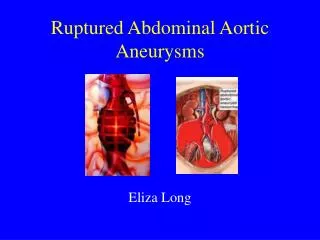
Abdominal Aortic Aneurysm • Older individual >60 yrs age • Sudden onset epigastric, abdominal back or flank pains – severe • Beware ‘renal colic’ in over 65 yr old • Fall or syncope • Vitals may be normal to start • Will generally appear ‘unwell’ • Pulsatile mass present only 60% time • Labs – Massive transfusion protocol • Imaging – Bedside USS • ED management – IV access x 2 • Minimal volume resuscitation • Emergency transport to TWH Vascular OT via ambulance
Summary • Consider pain character to decide if pain is visceral or parietal • Localise to quadrant to narrow diagnosis, but be aware of variation • Be aware of ability to be led astray by labs in RUQ pathology – get an USS (bedside) • Keep cholangitis in mind in your septic screen; appreciate it’s ability to deteriorate • Don’t get too concerned about pancreatitis scores in ED; if marked clinical or biochemical abnormality consider ICU • Dyspepsia is challenging! Be careful not to label too early. • AAA at SDMH requires rapid diagnosis and emergency transport