Download
1 / 42
420 likes | 669 Views
The World Congress of Neuroradiology Bologna, Italy, Oct 4-9 2010. HIPPOCAMPAL MR VOLUMETRIC STUDIES IN PAEDIATRIC CONTROL AND EPILEPSY GROUP. Win Mar @ Salmah J, Noorfizura A , Mohd Shafie A, Ahmad Helmy A K, Salmi A R* Naing L** Department of Radiology, Department of Paediatric*,

E N D
The World Congress of Neuroradiology Bologna, Italy, Oct 4-9 2010 HIPPOCAMPAL MR VOLUMETRIC STUDIES IN PAEDIATRIC CONTROL AND EPILEPSY GROUP Win Mar @ Salmah J, Noorfizura A , Mohd Shafie A, Ahmad Helmy A K, Salmi A R* Naing L** Department of Radiology, Department of Paediatric*, School of Medical Sciences, Universiti Sains Malaysia, 16150 Kubang Kerian, Kelantan, MALAYSIA. **PAPRSB3 Institute of Health Sciences, Universiti Brunei Darussalam XIX SYMPOSIUM NEURORADIOLOGICUM Wed 6 Oct 2010 15:00 - 15:15
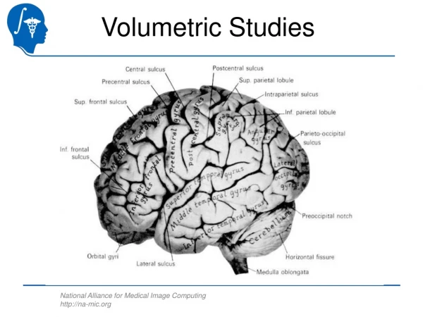
Introduction • Epilepsy is an important cause of neurological morbidity in children. • Cross et al., 1993, noted that hippocampal changes in children with intractable temporal lobe epilepsy were comparable to adults. • MRI features of hippocampus-morphology and volume – are part of the epilepsy imaging nowadays. • Visual analysis of MRI may detect gross hippocampal atrophy.
Introduction However, smaller degrees of volume loss may be overlooked and right and left asymmetry of size may be misinterpreted as volume loss. Side to side comparison of hippocampal volume can identify hippocampal sclerosis on the atrophic side, but absolute hippocampal volumes detect bilateral atrophy. Normative data of local area is important to assist better evaluation and decision.
Objectives General objectives- • normative data of the hippocampal volume in Malaysian paediatric population • hippocampal volume in paediatric epilepsy patients
Objectives The aims of this study are to determine the hippocampal volume in children with epilepsy to determine the hippocampal volume in children in the control group; to compare the hippocampal volume in children with epilepsy and the control group to compare the mean of right and left hippocampal volume in control subjects .
Material and methods This study was carried out in Universiti Sains Malaysia (USM) from January 2008 – June 2009. • This is a cross sectional study • 40 children with epilepsy and 40 children of normal control volunteer group. • Age = 7 to 18-years old • Malaysian • Approved by Ethics committee on 21 April 2008 with the reference number of USMKK/PPP/JEPeM [200.4.(1.4)].
Material and methods- subjects Epilepsy group • children diagnosed with epilepsy • from paediatric clinic and neurology clinic • Epilepsy was diagnosed based on clinical and EEG findings • Referred for neuroimaging study for epilepsy • Excluded • Organic lesion found on MRI
Material and methods-subjects Normal group • Children who are normal • Excluded • Epilepsy • Head injury • Psychiatric illnesses • Organic lesion found on MRI • from paediatric, radiology and neurology clinic • MRI performd after informed consents were obtained.
Material and methods- MR technique • MRI of brain and temporal lobe series • Signa Horizon LX 1.0 Tesla from the General Electric Company. • MRI Brain was done first • Axial T1, T2, FLAIR and T1 sagittal images • It is used as screening for presence of mass lesion or other abnormalities • Sagittal image is used as a guide to obtain perpendicular plane to temporal lobe.
Material and methods- MR technique • Temporal lobe series • coronal images perpendicular to the long axis of the temporal lobe. • IR, T2, FLAIR of coronal 4mm slice thickness with 1mm gap and • 3D SPGR coronal 2mm thickness,1mm gap • parameters: • T1 (TE/TR, 11/420) • T2 (TE/TR, 79.3/4020) • FLAIR (TE/TR, 147/9002) • IR (TE/TR, 15/2000)-all have FOV 20X20,NEX 2.0.
Figure ‑1 : MRI plane for evaluation of the hippocampus. Lines indicates the plane for temporal lobe series. These lines are perpendicular to the long axis of the hippocampus. It starts from the most anterior part of the temporal lobe, extends posteriorly to the splenium of corpus callosum
Material and methods-Anatomy • Hippocampal boundaries - based on protocol of Obenaus et al., 2001. • The whole hippocampal volume was measured. • Measurement of hippocampal body and tail included subicular complex, the hippocampus proper, the dentate gyrus, the alveus and hippocampal fimbria. • Measurement excluded the amygdala, parahippocampal gyrus, the isthmus of the cingulate gyrus and the crus of fornix.
Material and methods-volume measurement • manually • Osirix workstation (v 3.5.1-64 bit). • Right and left -measured individually. • ROI -manually traced with - computer mouse. • A value of the area appeared after completion. • measured three times for each ROI and the average was taken. • The same procedure was continued until the whole hippocampus was covered.
Material and methods-volume measurement • summed the average of each slice = the total area of hippocampus for each side. • Volumes = total area of the hippocampal complex (cm²) x the slice thickness (0.5cm) = volume of the hippocampal complex (cm³).
Figure 2 : Manual tracing of the hippocampal region This figure shows the outline border of the hippocampal region at the level of hippocampal body. The tracing of hippocampal body is shown as white line. It displays the total area in centimeter square (cm² ).
Material and methods-analysis • Data were analyzed by SPSS (version 14.0) • The H volumes of both control and epilepsy patients were analysed using independent t test. • For the entire test, p value of less than 0.05 (p<0.05) was taken as significant. Results were expressed as mean and standard deviation (SD).
ResultsSubject’s Characteristics Table 1. Demographic Characteristics a= Independent t test. b = Fisher’s exact Test
ResultsSubject’s Characteristics Table 2. Distribution of age and sex.
Results1.Hippocampal volume in epilepsy group Table 3. The hippocampal volumes for each age group
Discussion 1. The hippocampal volume in the epilepsy group • Our study - -Epilepsy group – • Right = 2.47 cm³ (SD 0.52) • Left = 2.40 cm³ (SD 0.44) • mean of both sides was 2.43 cm³ (SD 0.44) • Wu et al., 2005 • 18 months to 15 years old • overall mean H volume was 2.30 cm³ (SD 0.33) for temporal lobe epilepsy with febrile convulsion • 2.34 cm³(SD 0.33) temporal lobe epilepsy only group.
Discussion 1. The hippocampal volume in the epilepsy group • Daley et al., 2006 (European) • 4-14 years old • overall mean H volume was 3.10 cm³ (SD 0.10) for temporal lobe epilepsy with febrile convulsion • 2.34 cm³(SD 0.33) temporal lobe epilepsy only group.
Discussion 1. The hippocampal volume in the epilepsy group • Most of the studies - hippocampal volumetry were related to TLE. • In our epilepsy group (n=40), • EEG records were available (n=28) • 12 cases were of temporal lobe origin • 16 cases were non-temporal in origin. • 12 cases who did not have EEG done. • Overall- various types and origin of epilepsy. • This finding suggests that, various types of epilepsy other than temporal lobe epilepsy, also causes reduction in hippocampal size.
Discussion 1. The hippocampal volume in the epilepsy group H size reduction -the seizure is of extrahippocampal onset [EHO]. • Surges R et al., 2008 – • the human hippocampus was invariably invaded by epileptic activity - -during secondarily generalised tonic–clonic seizures of EHO. • The presence of spontaneous epileptic activity in the group with an extrahippocampal lesion revealed persistent modifications in hippocampal excitability. This might be due to secondary epileptogenesis in the hippocampus following repeated seizures of EHO.
Discussion 1. The hippocampal volume in the epilepsy group H size reduction –contralateral to focus • Varho et al., 2005 – • 11 children with medically controlled focal epilepsy and infrequent seizures, most of whom had secondarily generalized tonic clonic seizures. In these children, the hippocampi contralateral to the focus was also affected.
Discussion 1. The hippocampal volume in the epilepsy group H size reduction –contralateral to focus • Varho et al., 2005 – • 11 children with medically controlled focal epilepsy and infrequent seizures, most of whom had secondarily generalized tonic clonic seizures. In these children, the hippocampi contralateral to the focus was also affected. • Above studies –support our findings- H vol reduction –epilepsy foci of various origins
Results2. Hippocampal volume in control group Table 4. The hippocampal volumes for each age group
Discussion 2. The hippocampal volume in the control group • Our study control group – • Right = 2.81 cm³ (SD 0.38) • Left = 2.65 cm³ (SD 0.41) • mean of both sides was 2.73 cm³ (SD 0.38) • India (Mulani et al., 2005) • the mean right H volume = 2.75 cm³ • the mean left H volume = 2.49 cm³ • age group of 6 to 12 years old. • China (Wu et al., 2005). • 2.61 cm³ (SD 0.21) on both sides • for control group (n=16) • ages - 18 months to 15 years.
Discussion 2. The hippocampal volume in the control group • European paediatric age group where the mean hippocampal volume was 3.25 cm³ (SD 0.46) (Varho et al., 2005).
Discussion 2. The hippocampal volume in the control group • our study revealed - No age effect on the H volume in control group of 7 to 18 years -- • Giedd J et al., 1996 – • A developmental study= postmorterm & MRI measurement • H reaches its maximum size at the age of 2 to 3 years old and • no absolute changes (or a small increase) in the age range of 4-18 years old.
Discussion 2. The hippocampal volume in the control group No age effect on the H volume in control group of 7 to 18 years --our study revealed - • MacMaster et al.,, 2004 • Christoph P. et al., 2006 • age 13 to 18 years old healthy children and adolescents
Results3. Comparism between epilepsy and control group Table 5. Comparison mean hippocampal volume (cm³) a Average of Right and Left HV b Independent t test
Discussion 3. Comparison of the hippocampal volume in children with epilepsy and the control group • Our study had shown that the H volume in patients with epilepsy was significantly smaller than normal subjects on both sides (p=0.001) • Szabo et al., 1995 -patient with complex febrile seizure, • Varho et al., 2005 - children with non-symptomatic focal epilepsy. • The mean H volume of the focus side of patients was significantly reduced compared with that of the controls.
Results4. Comparism of Right and left H vol in control group Table 6: Comparison mean of right and left hippocampal volume (cm³)
Discussion 4. Comparison of mean of right and left hippocampal volume in control group • Our study -Right H volume was much greater than the left, with p <0.001. • H volume Right >Left • Niemann et al., 2000 • Tisserand et al., 2000 • Giedd et al., 1996 – in 99 healthy children and adolescents • Ho et al., 1998b - 8 healthy children, left H was 8% smaller
Conclusion • Our findings are similar to other studies, like H vol: • in normal control • epilepsy children- -are similar to other Asian studies • significant reduction of the hippocampal volume in epilepsy patients • larger hippocampal volume on right side in control group.
Conclusion • Our study revealed reduction in hippocampal size epilepsy children whose focus were from various origin including temporal lobe. • The mean hippocampal volume in the control group provides the normative data for Malaysian children aged between 7 to 18 years.
Acknowledgements The authors wish to express their gratitude to Universiti Sains Malaysia for supporting this study by granting the Research University Grant umber 1001/PSKBP/8120197
References • Bronen, R. A., Cheung, G., Charles, J. T., Kim, J. H., Spencer, D. D., Spencer, S. S., Sze, G. & McCarthy, G. (1991). Imaging findings in hippocampal sclerosis: correlation with pathology. Am J Neuroradiol,12 (5), 933-40. • Cheon, J. E., Chang, K. H., Kim, H. D., Han, M. H., Hong, S. H., Seong, S. O., Kim, I. O., Lee, S. G., Hwang, Y. S. & Kim, H. J. (1998). MR of hippocampal sclerosis: comparison of qualitative and quantitative assessments. Am J Neuroradiol,19 (3), 465-8. • Christoph P. Wiedenmayer, R. B., George M. Anderson, Hongtu Zhu, Jose Amat, Ronald Whiteman, and Bradley S. Peterson (2006). Cortisol levels and hippocampus volumes in Healthy Preadolescent Children. Biol Psychiatry,60 856-861. • Cross, J. H., Jackson, G. D., Neville, B. G., Connelly, A., Kirkham, F. J., Boyd, S. G., Pitt, M. C. & Gadian, D. G. (1993). Early detection of abnormalities in partial epilepsy using magnetic resonance. Arch Dis Child,69 (1), 104-9. • Daley, M., Ott, D., Blanton, R., Siddarth, P., Levitt, J., Mormino, E., Hojatkashani, C., Tenorio, R., Gurbani, S., Shields, W. D., Sankar, R., Toga, A. & Caplan, R. (2006). Hippocampal volume in childhood complex partial seizures. Epilepsy Res,72 (1), 57-66. • Giedd, J., AC, V. & SD, H. (1996). Quantitative MRI of the temporal lobe, amygdala, and hippocampus in normal human development : ages 4-18 years. J Comp Neurol,366 223-30. • Ho, S. S., Kuzniecky, R. I., Gilliam, F., Faught, E., Bebin, M. & Morawetz, R. (1998b). Congenital porencephaly: MR features and relationship to hippocampal sclerosis. AJNR Am J Neuroradiol,19 (1), 135-41.Jack, C. R., Jr. (1994). MRI-based hippocampal volume measurements in epilepsy. Epilepsia,35 Suppl 6 S21-9.
References • Ho, S. S., Kuzniecky, R. I., Gilliam, F., Faught, E., Bebin, M. & Morawetz, R. (1998b). Congenital porencephaly: MR features and relationship to hippocampal sclerosis. AJNR Am J Neuroradiol,19 (1), 135-41.Jack, C. R., Jr. (1994). MRI-based hippocampal volume measurements in epilepsy. Epilepsia,35 Suppl 6 S21-9. • Kalviainen, R., Salmenpera, T., Partanen, K., Vainio, P., Riekkinen, P., Sr. & Pitkanen, A. (1997). MRI volumetry and T2 relaxometry of the amygdala in newly diagnosed and chronic temporal lobe epilepsy. Epilepsy Res,28 (1), 39-50. • Lawson, J. A., Vogrin, S., Bleasel, A. F., Cook, M. J. & Bye, A. M. (2000b). Cerebral and cerebellar volume reduction in children with intractable epilepsy. Epilepsia,41 (11), 1456-62. • Lehericy, S., Baulac, M., Chiras, J., Pierot, L., Martin, N., Pillon, B., Deweer, B., Dubois, B. & Marsault, C. (1994). Amygdalohippocampal MR volume measurements in the early stages of Alzheimer disease. Am J Neuroradiol,15 (5), 929-37. • Lehericy, S., Semah, F., Hasboun, D., Dormont, D., Clemenceau, S., Granat, O., Marsault, C. & Baulac, M. (1997). Temporal lobe epilepsy with varying severity: MRI study of 222 patients. Neuroradiology,39 (11), 788-96. • MacMaster, F. P. & Kusumakar, V. (2004). Hippocampal volume in early onset depression. BMC Med,2 2. • Mulani, S. J., Kothare, S. V. & Patkar, D. P. (2005). Magnetic resonance volumetric analysis of hippocampi in children in the age group of 6-to-12 years: a pilot study. Neuroradiology,47 (7), 552-7.
References • Niemann, K., Hammers, A., Coenen, V. A., Thron, A. & Klosterkotter, J. (2000). Evidence of a smaller left hippocampus and left temporal horn in both patients with first episode schizophrenia and normal control subjects. Psychiatry Res,99 (2), 93-110. • Obenaus, A., Yong-Hing, C. J., Tong, K. A. & Sarty, G. E. (2001). A reliable method for measurement and normalization of pediatric hippocampal volumes. Pediatr Res,50 (1), 124-32. • Surges R, A Schulze-Bonhage, D-M Altenmüller (2008). Hippocampal involvement in secondarily generalised seizures of extrahippocampal origin. J Neurol Neurosurg Psychiatry ;79:924-929 doi:10.1136/jnnp • Tisserand, D. J., Visser, P. J., van Boxtel, M. P. & Jolles, J. (2000). The relation between global and limbic brain volumes on MRI and cognitive performance in healthy individuals across the age range. Neurobiol Aging,21 (4), 569-76. • Varho, T., Komu, M., Sonninen, P., Lahdetie, J. & Holopainen, I. E. (2005). Quantitative HMRS and MRI volumetry indicate neuronal damage in the hippocampus of children with focal epilepsy and infrequent seizures. Epilepsia,46 (5), 696-703. • Wu, W. C., Huang, C. C., Chung, H. W., Liou, M., Hsueh, C. J., Lee, C. S., Wu, M. L. & Chen, C. Y. (2005). Hippocampal alterations in children with temporal lobe epilepsy with or without a history of febrile convulsions: evaluations with MR volumetry and proton MR spectroscopy. Am J Neuroradiol,26 (5), 1270-5.