Download
1 / 34
340 likes | 356 Views
Learn about phase contrast imaging vs. conventional radiography, methods for achieving phase contrast, and results in breast imaging and other applications. Explore innovative approaches and preliminary results in mammography, cartilage and soft tissue imaging. Discover how phase retrieval techniques with synchrotron and conventional sources can provide detailed results with reduced radiation exposure.

E N D
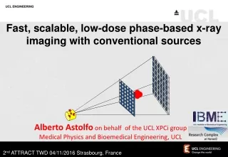
Fast, scalable, low-dose phase-based x-ray imaging with conventional sources Alberto Astolfo on behalf of the UCL XPCi group Medical Physics and Bioemedical Engineering, UCL 2nd ATTRACT TWD 04/11/2016 Strasbourg, France
Phase Contrast Imaging vs. Conventional Radiography Refractive index: n = 1 - di b; d>>b -> phase contrast (DI/I0~ 4pdDz/l) >> absorption contrast (DI/I0 ~ 4pbDz/l Two possible approaches: - detect interference patterns - detect angular deviations
Impressive results are achieved in breast imaging absorption phase contrast Arfelli et al. Phys. Med. Biol. 43 (1998) 2845-52
Free Space Propagation works wonders when implemented with a spatially coherent source – why ask for more? - It suffer immensely when transferred to conventional sources: the spread associated with projected source size becomes too large and kills the signal. Moreover: The system has little flexibility - only dsd can be changed But: Amazing stuff @ synchrotrons, e.g. check out Cloetens’ work at the ESRF + straightforward use e.g. coupled with Paganin’s single distance phase retrieval Olivo et al. Med. Phys. 28 (2001)1610-19
Other methods to perform phase contrast imaging: “Analyzer Based Imaging” Davis et al, Nature 373 (1995) 595-8; Ingal & Beliaevskaya, J. Phys. D 28 (1995) 2314-7, Chapman et al, Phys. Med. Biol. 42 (1997) 2015-25 - but even before that Forster 1980!
A different way to obtain a similar effect: The Edge Illumination Technique Provides results similar to ABI but opens the way to the use of divergent and polychromatic beams Olivo et al. Med. Phys. 28 (2001)1610-19
THE METHOD CAN BE ADAPTED TO A DIVERGENT AND POLYCHROMATIC (=conventional) SOURCE photons creating increased signal pre-sample apertured mask sample polychromatic, divergent beam (pre- shaping) detector pixels detail rotating anode x-ray source (focal spot 100 mm) photons creating reduced signal detector apertured mask Olivo and Speller Appl. Phys. Lett. 91 (2007) 074106
Little loss of signal intensity for source sizes up to 100 µm Which can be achieved with state-of-the-art mammo sources Why? Because we are only relying on refraction, which survives under relaxed coherence conditions; Because we are use aperture pitches matching the pixel size, i.e. BIG: the projected source size remains < pitch, and therefore blurring does “not” occur. Olivo and Speller Phys. Med. Biol. 52 (2007) 6555-73
Preliminary results - mammo (a): GE senographe Essential ADS 54.11; 25 kVp, 26 mAs (b): edge-illumination XPCi, 40 kVp, 25 mA – ENTRANCE dose 7 mGy (< mammo!) It has to be said the tissue was 2.5 cm thick -> we expect ~ same dose for thicker tissues Olivo et al Med. Phys. (letters) 40 (2013) 090701
Preliminary results - mammo (a): GE senographe Essential ADS 54.11; 25 kVp, 26 mAs (b): edge-illumination XPCi, 40 kVp, 25 mA – ENTRANCE dose 7 mGy (< mammo!) Tissue thickness 2 cm-> extrapolation leads to ~standard mammo dose for thicker (4-5 cm) tissues unpublished
Preliminary results - cartilage imaging Rat cartilage, ~ 100 µm thick, invisible to conventional x-rays Marenzana et al, Phys. Med. Biol. 57 (2012) 8173-84 under submission
Quantitative phase contrast imaging “SLOPE -” “SLOPE +” Titanium Aluminum PEEK Highly precise retrieval, for both high and low Z materials, up to high gradients where other methods break down Munro et al Opt. Exp. 21 (2013) 647-61
Phase retrieval with synchrotron and conventional sources: Ti filament: retrieved @ synchrotron and with conventional source! @ conventional source: incoherence modelled as beam spreading – the movement of the “spread” beam is then tracked and referred back to the phase shift that caused it. But with lots of care as far as “effective energy” is concerned! (See Munro & Olivo Phys. Rev. A 87 (2013) 053838) Munro et al, PNAS 109 (2012) 13922-7
preliminary CT results Soft tissue inside wasp thorax resolved Dose tens of mGy, instead of tens of Gy! Hagen et al, Med. Phys. (letters) 41 (2014) 070701
preliminary CT results Rabbit oesophagous (detergent enzymatic treatment – DET) Hagen et al, Sci. Rep. 5 (2015) 18156
preliminary CT results Rat Heart unpublished
Three-shot DARK FIELD IMAGING retrieval Endrizzi et al, Appl. Phys. Lett.104 (2014) 024106
Three-shot DARK FIELD IMAGING retrieval Endrizzi et al, Appl. Phys. Lett.104 (2014) 024106
DARK FIELD IMAGING of breast calcifications 3 images only, still within clinical dose limits! ENTRANCE dose 12 mGy (still compatible with mammo) Endrizzi et al, Appl. Phys. Lett.104 (2014) 024106
Microbubbles: a new concept of “phase-based” x-ray contrast agent Millard et al. Appl. Phys. Lett. 103 (2013) 114105
Microbubbles: a new concept of “phase-based” x-ray contrast agent absorption dark field bubbles no bubbles bubbles no bubbles Millard et al. Appl. Phys. Lett. 103 (2013) 114105
X-ray phase contrast microscope Endrizzi et al, Opt. Lett. 39 (2014) 3332-5
X-ray phase contrast microscope NB both pure “phase” objects (80 kVp were used) Endrizzi et al, Opt. Lett. 39 (2014) 3332-5
Asymmetric Edge Illumination Detector X-rays Mask 1 Mask 2 Detector The asymmetric Mask1 allows to retrieve attenuation, refraction and dark-field images during a single sample scan acquisition. X-rays Mask 2 Mask 1 1.5 m 0.5 m M. Endrizzi, Sci Rep 6, 25466 (2016)
detector mask 2 • X-Tek 160 kV Tungsten source 160 W • Mask 1: 15 cm x 1 cm • Mask 2: 20 cm x 1.5 cm • Source to mask 1: 1.5 m • Source to detector: 2 m • XCounter Dual-Energy detector Cd-Te • Pixel size: 100 µm • FOV up to: 16 cm x 50 cm • Energy tested: up to 100 kV mask 1 x-ray beam sample stage
Conclusions: Edge-illumination XPCi is a NON-INTERFEROMETRIC, INCOHERENT and QUANTITATIVE x-ray phase contrast method working with conventional sources which: allows the use of fully divergent, fully polychromatic x-ray sources with focal spots of up to at least 100 m - with no additional collimation/aperturing; the use of large apertures in thin gold layers (-> no angular filtration), low-absorbing graphite substrates, moderate misalignments between masks allowed achieving a reduction in the exposure times - although demanding medical applications require further developments. Most of all they keep the dose at acceptable levels. requires aperture pitches of the order of ~50-100 m - therefore making fabrication, alignment, FOV (masks are available up to 30 cm) and energy scale-up easier. has been described both by wave & geometrical optics (but for source sizes like the ones we use they give the same results) and robust phase retrieval was achieved. Translated “back” to a coherent source, it enables unprecedented phase sensitivity which opens the way to NEW, PREVIOUSLY INACCESSIBLE SCIENTIFIC APPLICATIONS
The state of men’s health is in crisis. Men experience worse longer-term health than women and die on average six years earlier. Prostate cancer rates will double in the next 15 years. Testicular cancer rates have already doubled in the last 50. Three quarters of suicides are men. Poor mental health leads to half a million men taking their own life every year. That’s one every minute. Our fathers, partners, brothers and friends are facing this health crisis and it’s not being talked about. We can’t afford to stay silent. Support the ‘Phase-contrast’ Movember Team: http://moteam.co/mo-on-phase Thank you for your attention (and donations)