Leishmaniasis
Leishmaniasis. David P. Humber Department of Life Sciences University of East London. Lecture Topics. The parasite and vector The life-cycle Clinical features Diagnosis Epidemiology Chemotherapy Vaccination. Introduction. Leishmaniasis Protozoal disease of mammals Largely zoonotic


Leishmaniasis
E N D
Presentation Transcript
Leishmaniasis David P. Humber Department of Life Sciences University of East London
Lecture Topics • The parasite and vector • The life-cycle • Clinical features • Diagnosis • Epidemiology • Chemotherapy • Vaccination
Introduction Leishmaniasis Protozoal disease of mammals Largely zoonotic 23+ pathogenic species Cutaneous leishmaniasis Visceral Leishmaniasis
Phylum Order Family Genus Sarcomastigophora Kinetoplastida Trypanosomatidae Leishmania The Parasite
Promasitogte Insect Motile Midgut Amastigote Mammalian stage Non-motile Intracellular Morphology Digenetic Life Cycle
Promastigote Amastigote Morphology Flagella Kinetoplast Golgi Nucleus Cytoskeleton
Kinetoplast Promastigote in Culture Nucleus
Speciation • Similar morphology • DNA bouyant density • Isoenzyme profiles - Zymodemes • Monoclonal antibodies • DNA hybridisation - PCR
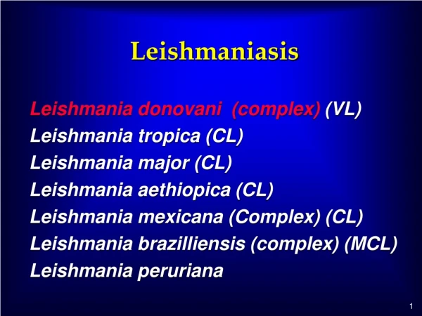
Species Pathogenic in Humans • Leishmania aethiopica • Leishmania brazilliensis (complex) • Leishmania donovani (complex) • Leishmania major • Leishmania mexicana (complex) • Leishmania tropica
Rodents Gerbils Hyraxes Bats Porcupines Opossums Sloths Primates Dogs Foxes Anteaters . . . . . Mammalian Hosts
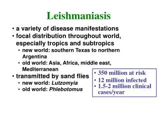
Vectors Phlebotomine Sandflies 6 genera world wide distribution Phlebotomus & Lutzomia 500 species Females Haematophagus Males sap feeders
Visceral Fatal (90% untreated) Liver Spleen Bone marrow Cutaneous Generally Self- healing Skin Mucous membranes Clinical Disease SPECTRUM OF DISEASE
Initial Infection • Similar in all species • Inoculation of promastigotes • Inflammation & chemotaxis • Receptor mediated phagocytosis Promastigote Amasitgote Transformation
Parasite Spread Macrophage lysis & parasite release Lymphatic spread Blood spread Target organs Skin/lymph nodes/spleen/liver/bone marrow
1903 1920 1931 William Leishman Pentavalent antimony Experimental transmission Visceral Leishmaniasis Leishmania donovani (complex) L.d. archibaldi - L.d.chagasi L.d.donovani - Ld.infantum
VL - Clinical Symptoms Variable - Incubation 3-100+ weeks Lowgrade fever Hepato-splenomegaly Bone marrow hyperplasia Leucopenia & Cachexia Hypergammaglobulinnemia
INFECTION Sub-clinical or inapparent infection RecoveryDeath Immune to reinfection Concurrent infection PKDL
Post Kala Azar Dermal Leishmanoid Normally develops <2 years after recovery Recrudescence Restricted to skin Rare but varies geographically
Diagnosis Clinical signs & symptoms Hypergammaglobulinemia ELISA/Formol gel Bone marrow biopsy Spleen or liver biopsy Culture & Histology
Marker L.. aethiopica L. tropica L. major L.. donovani Specificity of L. aethiopica primers
Treatment Good nursing & Diet Antibiotics Pentavalent antimony (upto 25% ressistance) Pentamidine Amidosidine New drugs - New delivery
Immune Response Innate IRs • Lsh/BCG gene • Lshr Lshs • No real human equivalent • Other species specific genes described • Complement • Polymorphs • Macrphages
Macrophages • Receptors • CR3 receptors for C3bi • Lipophosphoglycan • GP63 • Killing • Oxygen dependent • Oxygen independent
Macrophage activation • T cell activation • TH - 1 IL2, Gamma interferon • TH - 2 IL4, IL5 • SALT • Langerhans cells • Tissue dendritic cells
Vaccines • Leishmania + BCG • Ecuador - 3 species (Lbb,Lbg,Lma) • 2 doses killed whole parasites • 70% protection • Iran - 1 species (Lt) • 1 dose whole killed • 35% responded • 0% cf BCG alone
Old World Leishmania aethiopica Leishmania major Leishmania tropica New World Leishmania brazillensis L.b. L.b. Leishmania mexicana L.m L.m CL - Cutaneous Leishmaniasis Spectrum LCL - MCL - DCL
Localised Cutaneous Leishmaniasis • Single or multiple lesions • Usually on head and/or neck • Generally self-healing • Variable few week to many months • Ulceration followed by healing & scar • Secondary infection & tissue erosion
Mucocutaneous Leishmaniasis • Direct inoculation or extension • L.aethiopica & others • Low cell mediated immunity (CMI) • Metastatic spread • L.b.brazilliensis • High CMI & extensive tissue destruction • Also in DCL but no MI no tissue damage
Diffuse Cutaneous Leishmaniasis • Multiple diffuse spreading lesions • Usually face & limbs rarely trunk • No ulceration • Non-healing - life long infection • No cell mediated immunity • Good antibody response Leishmania aethiopica & Leishmania mexicana mexicana
Diagnosis Clinical feature & geographical location Skin biopsy/slit skin smear Culture & histology Monoclonal antibodies PCR
Treatment Control secondary infection Self-healing - probably no treatment Surgery/cryosurgery/Topical MCL & DCL Pentavalent antimony - pentamidine
Control • Vector control • Reservoir control • Treatment of active cases • Vaccination