Download
1 / 16
160 likes | 305 Views
Focusing X-rays to 1 nm – Requirements and Approaches. Jörg Maser Center for Nanoscale Materials X-ray Science Division NSLS II Workshop on Stability Apr. 19, 2007. Outline. Overall Considerations and Parameters Critical Specifications for Nanofocusing

E N D
Focusing X-rays to 1 nm – Requirements and Approaches Jörg Maser Center for Nanoscale Materials X-ray Science Division NSLS II Workshop on Stability Apr. 19, 2007
Outline • Overall Considerations and Parameters • Critical Specifications for Nanofocusing • Technical Approach to Instrument design • Short notes on Optics:
I) Overall considerations • Goal: reach a spatial resolution of 1 nm • Considerations: • What optics (not discussed here)? • How to measure? • Requirements for positioning/stability • Source • Beamline • Instrument • Assumptions: • Optics exists (distinguish monolithic and non-monolithic optics) • Diffraction-limited focusing required spatially coherent wavefront • No full-field optics feasible scanning for DAQ required
Typical nanofocusing parameters for NSLSII • Overall parameters of an NSLSII Nanofocusing Beamline (5 m straight) • Source distance: 50 m • Source size dv(FWHM) 3.8 um • Lateral coherence length at position of focusing optics (svq=l/p): 520 um, E = 10 keV • Focal length: 4.2 mm, E = 10 keV • Depth of focus: DOF = 20 nm Focusing Optics, Specimen Source
II) Source stability, size/position dl ~ /sin a ~ drN a • Size of measured focal spot: d = ( [dDL2 + (Msv)2 + Msource2]+ BL2+ instrument2) • M: Source magnification; : stability of subsystems • Example: • Multilayer Laue Lens as focusing optics: • drN = 0.9 nm • Msv = 0.28 nm, M = 710-5, M spatial coherence criterion (1/2, 1/p) • Dsource = 0.1 = 0.16 m, MDsource = 0.01 nm • Total size of focal spot: 0.94 nm (before beamline/instrument stability) spatially coherent wavefront
Source stability, angle • Coherent angle @ 10 keV: • q = 310-5 rad SR angle = 2/(g N) = 1.410-5 rad source divergence, v: y’ • No overfilling in vertical direction; angular fluctuations will directly lead to intensity variations • ’source = 0.1y’: I/I = 0.5%. • Required stability experiment-dependent • Spectroscopy, dichroism experiments very sensitive • Sufficient for fluorescence imaging • I could be addressed by XBPM in beamline
II) Critical Specifications for Nanofocusing – Overall considerations • How to measure? • Assume we have a suitable specimen << 1 nm features, thickness ~20 nm (t < DOF) • Assume we have an optimized detector/electronics • sr detection, 50% efficiency, high count rate capability: > 106 Hz 1 msec data acquisition time (based on CNM NPI simulations) • Assume 100 pixels for scans • Positioning requirement: • Require mechanical positioning of specimen/object of < 0.5 nm • Drift requirement: • Assume: B = 11021 Ph/s/mr2/mm2/0.1%BW • Line scan: require 1 pixel shift per scan line drifts < 4 nm/s • 2D scan: Require 10 pixel shift per line drifts < 4 Å/s
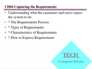
X-ray Fluorescence Imaging (Stefan Vogt, unpublished) Fig. 2.5: Simulation of fluorescence imaging of 5 nm thick test objects of Ti, Cu and W nanoparticles. The diameter of the test objects is 5 nm and 20 nm, respectively. An excitation energy of 12.2 keV is used. Fig. 2.5A and 2.5 B are for an exposure time of 15 msec. A solid angle acceptance of sr and a detector efficiency of 50% is assumed. The same simulation is shown in linear scale and log scale. Fig. 2.5 C shows the same object for an exposure time of 1.5 seconds (log scale). The position of individual nanoparticles of 5 nm diameters is indicated by blue arrows. A) Assumed: B = 51019 Ph/s/mr2/mm2/.1%BW Coherent Flux: Fcoh= 5109 Photons/s @ 12.2 keV B) C) From CNM Nanoprobe Instrument PDR, June, 2005
Consideration for beamline design • Nonsymmetric source size • Astigmatic beamline design required • Spatial filter remove source/BL optics motion effects • High position accuracy requirements for beam defining aperture Monochromator Optical Design CNM Nanoprobe Beamline BDA
Critical Components and relevant positioning specifications << 10-7 rad Instrument < 1.9 um Source • Lateral stability requirement: << 1 nm • Longitudinal requirement: << 20 nm • Monochromator angular stability (1st vs 2nd crystal): << 0.1 mrad, DCM at 25 m • Stability source vs instrument: << 3.8 um << 1 nm Monochromator << 20 nm
Other considerations • Required Thermal stability • Typical overall dimensions of optics/specimen assembly: 100 mm • Invar: = 1.610-6 m/m/K • expansion of 160 nm/K T < 2.510-3 K • However: significant are only differential drifts • Design using proper geometry can significantly reduce need for temperature stabilization • Tradeoff between temperature stability and air flow/acoustically excited vibrations
Sample EDS III) Technical approach to instrument design: • Overall considerations: • Amplitude of floor vibration: 20 – 30 nm • Provide nanometer positioning accuracy • Stiff design: Aim at in-phase “vibration” of sample and optics • Use of similar components/geometries for “stacks” of positioners • Position encoding and feedback • Conceptual Challenge: • Position sensors cannot be placed close to specimen/zone plate. Typical distance: 10 – 20 mm careful engineering of optics/specimen support
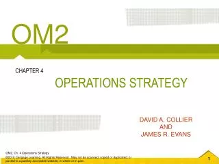
CNM Hard X-ray Nanoprobe Beamline at the Advanced Photon Source, 26-ID • Hard X-ray Nanoprobe Beamline Nanoprobe Instrument
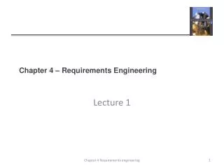
6 1 nm 4 2 0 20 10 Time [sec] Hard X-ray Nanoprobe Instrument • CNM/APS design; XRADIA TXM + engineering X-rays Sample Zone plate Flexure-based fine scanning stage Zone plate and specimen Module
Radius Thickness Some considerations on focusing optics Ideal Volume Optics • Desired: Monolithic Optics • Realistic 1 nm optics: non-monolithic approach might be necessary • e.g. 2-components optics: 12 degrees of freedom Currently fabricated at ANL 2D MLL
Summary • Source stability requirements – specifications sufficient for nonofocusing using fluorescence detection • Position stability: 0.1y • Source size stability 0.1y • Beamline stability • Monochromator: Dq << 0.1 mrad • Beamline apertures (e.g. h: D << 20 um). Heatload issues to be considererd • Instrument/source motion: < • Instrument design/stability • Instrument to source: D << • Optics/specimen stability: D << 1 nm (lateral), D < 20 nm (longitudinal)