Download
1 / 3
30 likes | 192 Views
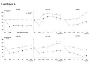
Pöschl et al., Suppl. Figure 1. a. Math1-cre::SmoM2 Fl/+ Ctnnb1(ex3) Fl/+. Math1-cre. Math1-cre::SmoM2 Fl/+. P0. EGL. IGL. EGL. P2. ML. PL. IGL. EGL. P7. ML. PL. IGL. b. Math1-cre::SmoM2 Fl/+ Ctnnb1(ex3) Fl/+. Math1-cre. Math1-cre::SmoM2 Fl/+. H&E. P16. Ki67. Ki67. Ki67.

E N D
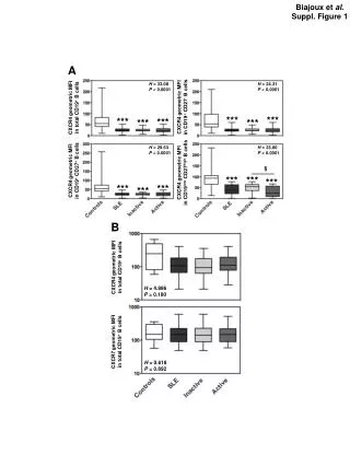
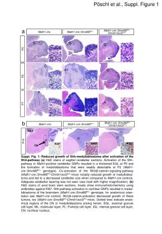
Pöschl et al., Suppl. Figure 1 a Math1-cre::SmoM2Fl/+ Ctnnb1(ex3)Fl/+ Math1-cre Math1-cre::SmoM2Fl/+ P0 EGL IGL EGL P2 ML PL IGL EGL P7 ML PL IGL b Math1-cre::SmoM2Fl/+ Ctnnb1(ex3)Fl/+ Math1-cre Math1-cre::SmoM2Fl/+ H&E P16 Ki67 Ki67 Ki67 CN Suppl. Fig. 1:Reduced growth of Shh-medulloblastoma after activation of the Wnt-pathway(a) H&E stains of sagittal cerebellar sections.Activation of the Shh-pathway in Math1-positive cerebellar GNPs resulted in a thickened EGL at P0 and the formation of medulloblastoma that were readily detectable at P2 (Math1-cre::SmoM2Fl/+ genotype). Co-activation of the Wnt/β-catenin-signaling-pathway (Math1-cre::SmoM2Fl/+Ctnnb1(ex3)Fl/+mice) notably reduced growth of medulloblas-toma and led to a decreased cerebellar size when compared to Math1-crecontrols. Adequate cerebellar layering was not seen (see inset with higher magnification). (b)H&E stains of axial brain stem sections. Insets show immunohistochemistry using antibodies against Ki67. Shh-pathway-activation in cochlear GNPs resulted in medul-loblastoma of the brainstem (Math1-cre::SmoM2Fl/+ genotype, for anatomical orien-tation see Math1-crecontrol). Wnt/β-catenin-activation decreased growth of these tumors, too (Math1-cre::SmoM2Fl/+Ctnnb1(ex3)Fl/+mice). Dotted lines indicate anato-mical regions of the CN or medulloblastoma arising herein. EGL: external granule cell layer, ML: molecular layer, PL: Purkinje cell layer, IGL: internal granule cell layer, CN: cochlear nucleus.
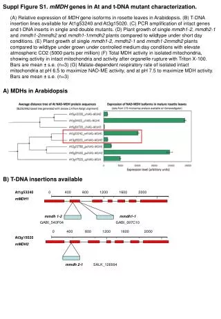
Pöschl et al., Suppl. Figure 2 c a b Wild type: Tumor DNA Tail DNA ** Exon 3 Exon 4 Exon 2 Marker 20 H2O A C B 370 bp ** 15 Wild type 715 bp Floxed: -catenin positive tumor cell nuclei [%] Floxed 10 loxP loxP Exon 2 Exon 3 Exon 4 Recombined 5 540 bp A B C Wild type Ca. 1 kb 0 Recombined: Exon 2 loxP Exon 4 P32 P16 P8 425 bp A d C IRES-GFP 1.5 Relative fraction of BrdUrd+ cells among GFP+ granule cells [%] Cre-IRES-GFP n. s. * 1 0.5 0 Math1-cre:: SmoM2Fl/+ Ctnnb1(ex3)Fl/+ Math1-cre:: SmoM2Fl/+ Suppl. Fig. 2: Incomplete recombination of the Ctnnb1allele.(a)The proportion of tumor cells with nuclear β-catenin-positivity decreased over time (P8 vs. P16: p=0.005, P8 vs. P32: p=0.001). (b)Scheme showing detectable PCR-fragments depending on allelic sequence. A, B and C delineate primers that were used for the analyses shown in (c). (c)The gel electrophoresis after PCR demonstrates incomplete recombination of the floxedCtnnb1(ex3) allele in tumor tissue of Math1-cre::SmoM2Fl/+Ctnnb1(ex3)Fl/+mice. (d)Primary cell cultures of Math1-cre::SmoM2Fl/+andMath1-cre::SmoM2Fl/+Ctnnb1(ex3)Fl/+tumors were transduced with Cre-IRES-GFP virus to allow recombination of yet unrecombined alleles. While Math1-cre::SmoM2Fl/+tumor cells displayed no difference in proliferation, Math1-cre::SmoM2Fl/+Ctnnb1(ex3)Fl/+tumor cellsshowed a significant decreaseof proliferation 24 h after transduction (p=0.023. Values were normalized to the mean of control treated cells of each genotype, respectively (value was set 1). n. s.: not significant, one asterisk: p<0.05, two asterisks p<0.01.
Pöschl et al., Suppl. Figure 3 b a c hGFAP-cre::SmoM2Fl/+ Ctnnb1(ex3)Fl/+ hGFAP-cre::SmoM2Fl/+ hGFAP-cre H&E H&E H&E H&E β-catenin Ki67 H&E β-catenin Ki67 H&E β-catenin Ki67 Suppl. Fig. 3: Impaired proliferation in hGFAP-positive precursors with Shh after the additional activation of Wnt/β-catenin-signaling.(a, b, c) Sagittal sections of hGFAP-cre(a), hGFAP-cre::SmoM2Fl/+(b) and hGFAP-cre::Ctnnb1(ex3)Fl/+SmoM2Fl/+(c) cerebella at P0. Higher magnifications of the external granule cell layer (EGL) of respective genotypes show immunohistochemistry using antibodies against β-catenin and Ki67. Shh-pathway-activation in hGFAP-positive precursor cells resulted in the formation of medulloblastoma that arose from the EGL and displayed a high proliferation rate (b). Constitutive co-activation Wnt/β-catenin-signaling in hGFAP-cre::Ctnnb1(ex3)Fl/+SmoM2Fl/+ led to a dramatically thinned EGL showing decreased proliferation, even compared to the control (a). Adequate cerebellar layering was not seen (c versus a). Nuclear positivity for β-catenin was detectable within the EGL (white arrow in the inset of c).