Download
1 / 26
260 likes | 339 Views
Explore the functions and parts of the respiratory system, from the nasal cavity to the lungs, to understand how breathing works.

E N D
INTRODUCTION The respiratory system generally includes tubes, such as the bronchi, which are used to carry air to the lungs where gas exchange occurs. The diaphragm, like other muscles can contract and relax. When someone inhales, the diaphragm contracts and flattens and the chest cavity expands. This contraction creates a vacuum that sucks air into the lungs. When exhaling, the diaphragm relaxes and returns to its previous position (dome-like shape) and the air is expelled from the lungs.
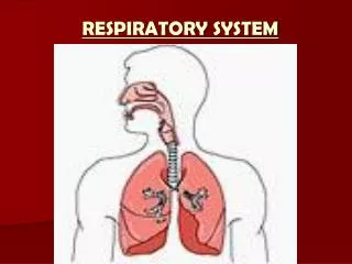
PARTS OF THE RESPIRATORY SYSTEM • Nasal Cavity • Epiglottis • Pharynx • Larynx • Trachea • Lungs • Diaphragm
NASAL CAVITY • The nostrils or nares are usually locatedin the openings of the nose in some animals. They open to the outside and let air enter the body.
EPIGLOTTIS • The epiglottis is a cartilaginous structure valve that covers the entry of the larynx and moves up and down, preventing food from entering it and the trachea. It also marks the limit between the oropharynx and the laringopharynx. When breathing, air passes through the pharynx and goes towards the larynx while the epiglottis remains opened. • The epiglottis closes when food gets swallowed and goes down the pharynx. This prevents food from obstructing the larynx.
EPIGLOTTIS • The epiglottis guards the entrance of the glottis, the opening between the vocal folds. It is normally pointed upward during breathing with its underside functioning as part of the pharynx, but during swallowing, elevation of the hyoid bone draws the larynx upward; as a result, the epiglottis folds down to a more horizontal position, with its upper side functioning as part of the pharynx. In this manner it prevents food from going into the trachea and instead directs it to the esophagus, which is further down the throat.
PHARYNX • The human pharynx is the part of the throat situated immediately behind the mouth and the nasal cavity, and above the oesophagus, the larynx, and the trachea. The human pharynx is conventionally divided into three sections: the nasopharynx , the oropharynx, and the laryngopharynx. The pharynx is part of both the digestive and the respiratory systems, and it plays an important role in vocalisation.
LARYNX • The larynx, commonly called the voice box, is an organ in the neck of mammals involved in protecting the trachea against food aspiration, breathing and sound production. It manipulates pitch and volume. The larynx houses the vocal folds which are an essential component of phonation. The vocal folds are located just below, where the tract of the pharynx splits into the trachea and the oesophagus.
TRACHEA • The trachea (or windpipe) is the bony tube that connects the nose and mouth to the lungs. It is an important part of the vertebrate respiratory system: when an individual breathes, air that is caught flows into the lungs through the windpipe.
LUNGS • The lungs are the essential breathing organs in many animals, including most tetrapods, a group of fish, and snails. In mammals and more complex life forms, the two lungs are located in the chest on either side of the heart. Their principal function is to transport oxygen from the air into the bloodstream and release carbon dioxide from the bloodstream into the air. This exchange of gases is accomplished by the mosaic of specialised cells that form millions of tiny, exceptionally thin-walled air sacs called alveoli.
LUNGS To explain the anatomy of the lungs, it is necessary to discuss the passage of air through the mouth to the alveoli. Once air progresses through the mouth or the nose, it travels through the oropharynx, the nasopharynx, the larynx, the trachea, and a progressively subdividing system of bronchi and bronchioles, until it finally reaches the alveoli where the gas exchange of carbon dioxide and oxygen takes place.
DIAPHRAGM • It is a dome-shaped muscle that separates the chest cavity and the abdominal cavity. • It is characteristic of all mammals, and in birds it appears in a rudimentary way.
INHALATION • Inhalation is initiated by the diaphragm and supported by the external intercostalmuscles. Normal respirations are 10 to 18 breaths per minute,with each lasting around 2 seconds. During vigorous inhalation (at rates exceeding 35 breaths per minute), or when approaching respiratory failure, accessory muscles of respiration are recruited for support. These consist of the sternocleidomastoid and platysma muscles, and the scalene muscles of the neck. Pectoral muscles and latissimus dorsi are also accessory muscles.
INHALATION • Under normal conditions, the diaphragm is the primary driver of inhalation. When the diaphragm contracts, the ribcage expands and the contents of the abdomen are moved downward. This results in a larger thoracic volume and negative pressure (with respect to atmospheric pressure) inside the thorax. As the pressure in the chest falls, air goes into the conducting zone. Here, the air is filtered, warmed, and humidified as it flows to the lungs. • During forced inhalation, as when taking a deep breath, the external intercostal muscles and accessory muscles aid in further expanding the thoracic cavity.
EXHALATION • Exhalation is generally a passive process; however, active or forced exhalation is achieved by the abdominal and the internal intercostal muscles. During this process air is forced or exhaled out. • The lungs have a natural elasticity: as they recoil from the stretch of inhalation, air flows back out until the pressures in the chest and the atmosphere reach equilibrium. • During forced exhalation, as when blowing out a candle, expiratory muscles including the abdominal muscles and internal intercostal muscles generate abdominal and thoracic pressure, which forces air out of the lungs. • http://kidshealth.org/kid/htbw/RSmovie.html • http://www.lung.org/your-lungs/how-lungs-work/?gclid=CLTOgIaol74CFQKhOgodPnQA6Q
HEALTHY HABITS FOR CARING FOR OUR RESPIRATORY SYSTEM Exercise regularly, this helps to improve the circulation of the blood. Eat a well balanced diet. Eat vegetables, especially green vegetables. Avoid eating too much saturated fats. Use beneficial fats and oils. Maintain a healthy weight. Live in a clean environment. Avoid smoking cigarettes and second hand smoke. Smoking increases the risk of stroke and coronary heart disease. Have a positive outlook in life. Try to reduce stress and tension. Avoid high blood pressure because this can cause heart failure and stroke.
DISEASES OF THE RESPIRATORY SYSTEM Respiratory diseases can be classified in many different ways: by the organ involved, by the pattern of symptoms or by the cause of the disease. The main thing is to always be careful around food because some foods can cause allergic reactions and incite breathing difficulties. Some common examples include seafood (like prawns), some fatty fish, radish, arrow root, fish fingers, lemon, dhal, peanuts (dry fruits in general), water content spinach, curd, bananas, grapes, pomegranates, berries, custard apple, ice creams, etc. In the summer, bad weather conditions mean sandy and dusty weather or for some people, bad weather may be affect them in winter also.
Now you get to do research. • In table teams you will research and analyze diseases of the respiratory system. The following slides will get you started. Here is the break down of the table assignments: • Table 1 – INFLAMATORY LUNG DISEASE • Table 2 – ASTHMA • Table 3 – RESPIRATORY TRACT INFECTION • Table 4 – RESPIRATORY TUMOURS • Table 5 – PLEURAL CAVITY DISEASES • Table 6 – PULMONAR VASCULAR DISEASE • Criteria: • The presentation must have the following elements/information: • Respiratory system according to table • Cause of disease • Symptoms • Prognosis (outlook) • How does it damage the organ(s) • Treatment/Cure • At least three pictures
INFLAMATORY LUNG DISEASETable 1 Inflammatory lung disease is also called Chronic Obstructive Pulmonary Disease (COPD) and includes a wide range of inflammatory lung ailments. These ailments include asthma, emphysema and chronic bronchitis. In many cases, the lungs are chronically inflamed, making it difficult to breathe and placing strain on the heart. Individuals with inflammatory lung disease may find it difficult to engage in exercise or activities that require heavy breathing. COPD is an illness that is gaining in frequency as pollution in our air increases.
ASTHMATable 2 Asthma is a common chronic inflammatory disease of the airways characterized by variable and recurring symptoms, reversible airflow obstruction, and bronchospasm. Symptoms include wheezing, coughing, chest tightness, and shortness of breath. Treatment of acute symptoms is usually with an inhaled short-acting beta-2 agonist . Symptoms can be prevented by avoiding triggers, such as allergens and irritants, and by inhaling corticosteroids. Leukotriene antagonists are less effective than corticosteroids and thus less preferred. The prevalence of asthma has increased significantly since the 1970s. As of 2009, 300 million people were affected worldwide. In 2009 asthma caused 250,000 deaths globally. Despite this, with proper control of asthma with step down therapy, prognosis is generally good.
RESPIRATORY TRACT INFECTIONTable3 Respiratory tract infections affect the nose, the throat, and the airways, and may be caused by any of several different viruses. Common respiratory tract infections include the common cold and influenza (flu). Typical symptoms include nasal congestion, a runny nose, scratchy throat, cough, and irritability. The diagnosis is based on symptoms. Good hygiene is the best way to prevent these infections, and routine vaccination can prevent influenza. Treatment aims to relieve symptoms (palliative effect). There are two types: Lower and Upper respiratory tract infections.
RESPIRATORY TUMOURSTable 4 Malignant tumours Malignant tumours, or cancers of the respiratory system, particularly lung cancers, are a major health problem responsible for 15% of all cancer diagnoses and 29% of all cancer deaths. The majority of respiratory system cancers are attributable to smoking tobacco. In addition, since many cancers spread via the bloodstream and the entire cardiac output passes through the lungs, it is common for cancer metastases to occur within the lung. Breast cancer may invade directly through local spread, and through lymph node metastases. After metastasis to the liver, colon cancer frequently metastasizes to the lung. Prostate cancer, germ cell cancer and renal cell carcinoma may also metastasize to the lung.
Treatment of respiratory system cancer depends on the type of cancer. Surgery , chemotherapy and radiotherapy are all used. The chance of surviving lung cancer depends on the cancer stage at the time the cancer is diagnosed and is only about 14-17% overall. In the case of metastases to the lung, treatment can occasionally be curative but only in certain, rare circumstances. Benign tumours Benign tumours are relatively rare causes of respiratory disease. Examples of benign tumours are: Pulmonary hamartoma. Congenital malformations such as pulmonary sequestration and congenital cystic adenomatoid malformation.
PLEURAL CAVITY DISEASESTable 5 Pleural cavity diseases include emphysema and mesothelioma which are mentioned above. A collection of fluid in the pleural cavity is known as a pleural effusion. This may be due to fluid shifting from the bloodstream into the pleural cavity due to conditions such as congestive heart failure and cirrhosis. It may also be due to inflammation of the pleura itself as can occur with infection, pulmonary embolus, tuberculosis, mesothelioma and other conditions. A pneumothorax is a hole in the pleura covering the lung, allowing air in the lung to escape into the pleural cavity. The affected lung “collapses” like a deflated balloon. A tension pneumothorax is a particularly severe form of this condition where the air in the pleural cavity cannot escape, so the pneumothorax keeps getting bigger until it compresses the heart and blood vessels, leading to a life threatening situation.
PULMONAR VASCULAR DISEASETable 6 Pulmonary vascular diseases are conditions that affect the pulmonary circulation. Pulmonary embolism, a blood clot that forms in a vein, breaks free, travels through the heart and lodges in the lungs. Large pulmonary emboli are fatal, causing sudden death. A number of other substances can also clog the lungs but they are much more rare: fat embolism, amniotic fluid embolism , air embolism (iatrogenic - caused by invasive medical procedures).
Pulmonary arterial hypertension: elevated pressure in the pulmonary arteries. It is most commonlyidiopathic (i.e. of unknown cause) but it can be due to the effects of another disease, particularly COPD. This can lead to a strain on the right side of the heart, a condition known as cor pulmonale. Pulmonary edema: leakage of fluid from capillaries of the lung into the alveoli (or air spaces). It is usually due to congestive heart failure. Pulmonary hemorrhage: inflammation and damage to capillaries in the lung resulting in blood leaking into the alveoli. This may cause blood to be coughed up. A pulmonary hemorrhage can be due to auto-immune disorders such as Wegener's Granulomatosis and Goodpasture's syndrome.


