Download
1 / 55
650 likes | 949 Views
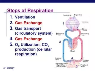
Mechanics of respiration. By Dr. M.B.Bhat. Mechanics of Respiration. Ventilation –Breathing in & out of air from lungs Inspiration –Taking in of air –Active process Expiration –Given out of air –Passive process (normally) –Active process in forced expiration Normal respiration by

E N D
Mechanics of respiration By Dr. M.B.Bhat.
Mechanics of Respiration • Ventilation –Breathing in & out of air from lungs • Inspiration –Taking in of air –Active process • Expiration –Given out of air –Passive process (normally) –Active process in forced expiration • Normal respiration by • Negative pressure breathing – • Advantage –increase venous return
Normal Respiration • During inspiration – • By operation of respiratory pump (Muscle Force)—Thorax expand –along with Lungs also expand– • Decreases pressure in thoracic cavity • By over-coming resistance • Rushing of air from atmosphere through the respiratory tract into lungs • Till intra-pulmonary & atmospheric pressure equals
During expiration – (cessation of inspiratory activity) By elastic recoiling – Thorax assumes its original position –Lungs get compressed – Thoracic cavity pressure rises – Air goes out (through the same routes) –till the air pressure between intra-pulmonary & atmospheric are equal Hence normal expiration is apassive process
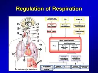
Mechanism of respiration • Involves 3 processes • Creation ofForce (for operation of respiratory pump) –by respiratory muscles • Pressure changes (in the thoracic cavities) • Resistance to over come (for air movements)
Respiratory muscles –Inspiratory & Expiratory muscles • Inspiratory muscles – • Chief inspiratory muscles (operate during quite respiration) –Diaphragm & External inter-costal muscles • Expiratory muscles –(operate only during forceful expiration) –Internal inter-costal muscles; Abdominal muscles • Accessory respiratory muscles (operate only in forceful respiration) –Scaleni, Sternocledo-mastoid, Anterior Serati, Extensor muscles of vertebral columns
Nerve supply –Phrenic nerve (C3,4&5) Contraction of diaphragm flatten it towards abdomen by 1.5cm (during quite respiration) & up to 7cm (during forceful respiration) Result –increase in vertical dimension of thoracic cavity Account for 75% change in intrathoracic volume during quite respiration (2/3rd of Tidal volume) Though important for respiration not absolute necessity
External inter-costalmuscles • Origin –From the lower border of one rib to -- • Insertion— upper border of the rib below • Course –From origin pass obliquely forwards & downwards to the lower rib • Nerve supply –Nerve to inter-costal muscles (T1 to T12) • Contraction –leads to upward & forward movements of ribs
Thoracic cavity • Thoracic lid –made up of 1st pair of ribs jointed between vertebral column & manubrium sterni –moves very little (in hyperpnea manubrium moves upwards –thereby increase antero-posterior diameter) • Upper costal area –2nd to 6th ribs –slope obliquely downward & forward from posterior to anterior • Lower costal area –7th to 10th ribs –swing outward & upwards
As Thoracic lid is immovable – Contraction external inter-costal muscles pull the lower ribs causing upward & outward movements Upward movements push manubrium upwards –increase antero-posterior diameter –Pump-handle movement Outward movements –by virtue of the bowed mid-part of ribs –increase transversediameter –Bucket-handle movement
By the contraction of diaphragm— Vertical dimension of thoracic cavity increases – to about 1.5cm during quite respiration & 7cm during forceful respiration By the action of inter-costal muscles & accessory muscles – Circumference of chest increases by – 1cm in quite respiration 5 to 11cm in forced respiration
Expiration isa quiet passivemotion Cessation of inspiratory muscle activity – Due to elastic recoiling tendency of lungs & chest –expiration takes place For forceful expiration – Internal inter-costal muscles (arrangement of which is quiet opposite to external inter-costal muscles) draws the ribs inwards & downwards Abdominal muscles –increase intra abdominal pressure and abdominal contents push diaphragm upwards; & draws the lower ribs downwards & medially thereby decrease thoracic dimension
Pressures of the thoracic cage • Two types of pressures • Intra-Pleural pressure (Or) Intra-Thoracic pressure (Pressure between two pleura) 2Intra-Alveolar pressure (Or) Intra-Pulmonary pressure (Pressure in the lungs –alveoli)
Intra pleural pressure (or) Intra thoracic pressure – • Normal value—During quite respiration • At the beginning of inspiration= –3 cm H2O (–2mm Hg) • At the end of inspiration= –7.5 cm H2O (–6mmg) • Max. in forced inspiration = –20 mm Hg • Ma. In forced expiration = +40 mm Hg
Measurement of intra pleural pressure • Direct-- • Indirect– Principle --pressure at lower 3rd of esophagus beneath the lungs is same as intra thoracic pressure –as esophagus also present in the thoracic cavity. • Procedure - Catheter tipped with a balloon is swallowed & kept at lower 3rd of esophagus and inflate & connect the catheter with manometer. • Reasons for negative pressure (explain)
Significance of intra pleural pressure • Always negative (sub-atmospheric) in normal respiration • Prevents collapse of alveoli • Prevents collapse of small air ways • Aids in venous return (Respiratory pump) • In Pneumothorax it becomes equal to atmospheric pressure –alveoli collapse
Intra alveolar pressure (or) Intra pulmonary pressure • At the beginning of inspiration & at the end of expiration –it is zero • During inspiration= minus 1to 4 mm Hg • During expiration – Plus 1to 4 mm Hg • During forced inspiration – minus 20 mm Hg • During forced expiration – plus 40 mm Hg • Measurement – “Mouth pressure”
Trans-pulmonary pressure • Difference between intra thoracic pressure & intra pulmonary pressure • Pressure operating inner & outer wall of the alveoli • Measure the elastic forces in the lungs
Resistances • Elastic resistance & • Non-elastic resistances –are; • Air-way resistance • Non-elastic tissue resistance
Elastic resistance • Resistance due to elastic nature of lungs & thoracic cage • Reciprocal of elastic resistance is compliance
Compliance • Compliance is the measure of stretch ability or elasticity • StaCompliance is change in volume by unit change in pressure (∆V/∆P) – L/cm H2O • Specific compliance –Compliance/FRC (in L per cm H2O per L)
Types of compliances • As both lungs & thoracic cage has elastic nature –in the respiratory system the various compliances are; • Lung compliance (L.C) • Thoracic compliance (Th.C) • Total compliance (T.C) -- (both L.C & Th.C) • Compliance is measured in static condition
Lung compliance • Volume of lung expansion for each unit increase in intra pleural pressure • Normal value –0.22L / cm H2O • (range – 0.09 to 0.26 / cm H2O) • In infant – 0.005 / cm H2O
Factors affecting lung compliance • Size • Phases of Respiratory cycle – Deflation & Inflation • Gravity –less at apices • Factors Compliance -- • Emphysema & Old age • Factors Compliance -- • Pul. Congestion, Pul. edema & fibrosis --
Total compliance • Normal value – 0.11L / Cm H2O • Measurement -- ∆V / ∆P (Airway pressure –Intra pulmonary Pressure)
Thoracic compliance • Is due to Sterno-costal joints • Normal value – 0.22L / cm H2O • Cannot measure directly • Calculate by using the equation-
Factors determining lung compliance • Elastic nature of pulmonary tissue (contribute 1/3rd) • Surface tension (contribute 2/3rd)
Elastic nature of pulmonary tissue • Inter woven of elastin & collagen fibers like nylon stocking arrangement • In emphysema –degradation of elastin & collagen frame work –leads to increase distensiblity • In old age –change in physico-chemical properties of elastin & collagen –increase distensibility • In pul.fibrosis –increase in interstitial tissue –stiffness – distensibility
Surface tension • Of pure water =70dynes/cm • Of Alveolar fluid without surfactant =50dynes/cm • Of Alveolar fluid with surfactant =5 dynes/cm • Laplace law – P = 2T/r
Average size alveoli (100µm) – the collapsing pressure Or distending pressure (P)-- • with surfactant; P= 4cm H2O (3 mm Hg) • Without surfactant; P = 18cm H2O
Surfactant • Surface tension lowering agent • Secreted from Type II cells • Contains – • Dipalmitoyl phosphatidyl choline (DPPC) (Dipalmitoyl lecithin) – 62% • Phosphatidly glycerol –5% • Other phospholipids –10% • Neutral lipids –13% • Protein – 8% • Carbohydrate – 2% • And calcium ions
Mechanism of action of Surfactant • Surfactant consist of mainly of–Phospholipids • Phospholipids have hydrophilic (phosphate portion) & hydrophobic (lipid portion) ends • The hydrophilic end dissolves into the alveolar fluid; while the hydrophobic end facing the exterior. • As the hydrophobic portion (lipid) has attraction towards gas, the inward attraction of the molecules of the surface area can be minimized & thereby lowering the surface tension.
Significance of surfactant • Surface tension (by 1/12th –1/2) • Compliance • Respiratory work load is • Helps in stability of alveoli of unequal size • Prevent collapse of alveoli during expiration • Prevent bursting of alveoli during inspiration • Keeps the alveoli dry (prevent pul. Congestion)
Factors affecting surfactant • Factors surfactant – • Occlusion of main bronchus • Occlusion of pulmonary artery • Long-term inhalation of 100% O2 • Cutting of both vagi • Cigarette smoking Factors surfactant – • Thyroid hormone -- production • Glucocorticoids – accelerate maturation
Clinical significance of surfactant • (Infant) Respiratory distress syndrome (IRDS) or • Hyaline membrane disease (due to deficiency of surfactant in fetal life)
Pulmonary Resistance • Non-elastic tissue resistance (Viscous resistance) • Air-way resistance Resistance are measured during dynamic condition R = ∆P/ V (flow) in liter/sec
Pulmonary resistance (Total resistance) • Air way resistance + viscous resistance • Obtained by using intra pleural pressure • 3.5cm H2O per liter per second
Air way resistance • Due to friction between molecules of flowing gas & also with walls of the tube • Raw = (Pmouth – Palv) / V in L/sec • Normal value = 0.6 to 2.4 cm H2O per Liter per second • LFTs used in determine Raw are – • PEFR; MVV; FEV • Raw --Increases in obstructive type respiratory diseases
Factors determining air way resistance Factors air wayresistance Decrease lung volume Decrease in diameter Total cross sectional area Increase in Density & viscosity Types of flow -- Dynamic compression of air ways
Non-elastic tissue resistance • Resistance offered by the non-elastic tissue of the thoracic cavity –Lungs, rib-cage, diaphragm & abdominal content • Responsible for the Hysteresis (closed loop) type of pressure-volume relation of respiratory cycle • Account for 20% of total pulmonary resistance • Itis increased greatly in Emphysema
Thoracic resistance • Pulmonary resistance (Airway resistance + viscous resistance) + chest wall resistance • Thoracic cage resistance –no measurement developed so far
Work done of breathing • Work done = Pressure x volume • Normal value of total work done = 0.3 to 0.8 kg m/min • Elastic work –65% • Non-elastic work –35%; out of this • Airway resistance work –28% • Viscous resistance work (Inertia work)–7% • Work of breathing increases in – • Emphysema, asthma, congestive cardiac failure with dyspnea & orthopnea