Download

1 / 12
120 likes | 325 Views
Chapter 8 Study Guide: The Nervous System. Anatomy Bowl Prep. Organs and Divisions of the Nervous System (understand). Central Nervous System (CNS) Brain Spinal Cord Peripheral Nervous System (PNS) Cranial Nerves Spinal Nerves Autonomic Nervous System (ANS): Involuntary Sympathetic

E N D
Chapter 8 Study Guide:The Nervous System Anatomy Bowl Prep
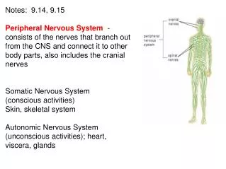
Organs and Divisions of the Nervous System (understand) • Central Nervous System (CNS) • Brain • Spinal Cord • Peripheral Nervous System (PNS) • Cranial Nerves • Spinal Nerves • Autonomic Nervous System (ANS): Involuntary • Sympathetic • Parasympathetic
CNS vs. PNS • Two types of cells found in the nervous system: • Neurons and glial cells • Neurons conduct impulses, whereas glial cells are for support • Each neuron consist of a cell body, dendrites, and an axon
Nerves • A nerve is a group of peripheral nerve axons bundled together like the strands of a cable • Because nerves usually have myelin sheath and myelin is white, nerves are called white matter in the PNS • Bundles of axons are called tracts, and may be myelinated and thus form this system of white matter • Dendrites is called gray matter because of its characteristic grey appearance • Understand that myelinated nerves have faster conduction velocity • Which means that they are able to carry a signal much faster than nonmyelinated fibers
Reflex Arc (understand concept) • Brain is not “consciously” involved: pathway of nerve fibers and does not ascend to communicate with the neurons of the brain after stimulus is applied. • Sensory (afferent) impulse • Sensory neuron cell bodies housed in the Dorsal Root Ganglion (DRG) • Motor (efferent) impulse • Motor neuron cell bodies housed in the Ventral (Anterior) Horn of the Gray Matter of Spinal Cord • Interneurons may be involved if the reflex arc is “polysynaptic” • Polysynaptic vs. Monosynaptic
Neurotransmission (understand) • Propagated action potential (AP) causes release of a neurotransmitter (NT) • Over 30 NTs identified • NTs are docked in vesicles at the axon terminus • NTs are released into the synaptic cleft • Bind to complementary receptors • Binding is either stimulatory or inhibitory to post-synaptic cell • Post-synaptic cell is either an effector cell (skeletal muscle or glandular cell) or another neuron
Divisions of the Brain(structure and function) • Cerebrum • Diencephalon • Thalamus • Hypothalamus • Cerebellum • Brainstem • Midbrain • Pons • Medulla Oblongata
Spinal Cord(structure and function) • Don’t worry about different spinothalamic tracts!!! • Understand that grey mater is found in the center of the spinal cord • White mater is found around the outer portion of the spinal cord • Meninges: are protective tissue layers that enclose the brain and spinal cord. The three layers are • Dura • Arachnoid • Pia • There are spaces in between the meningeal layers. • Ex: epidural layer, sub dural layer
Clinical Applications • Herpes Zoster or Shingles • Is a unique viral infection that almost always affects the skin of a single dermatome • A dermatome is a pathway that a nerve travels to innervate the skin • The result is that a patient would experience a painful eruption of red, swollen, plaques of vesicles on their skin