Download
1 / 36
360 likes | 379 Views
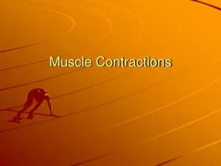
This text explains the different types of muscle contractions - summation, twitch, and tetanus. It also outlines the sliding filament model of muscular contraction and the role of ATP in muscle contraction.

E N D
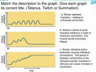
Match the description to the graph. Give each graph its correct title. (Tetanus, Twitch or Summation) a. Shows repeated impulses – leading to enhanced contraction. b. Shows a series of quick impulses leading to a state of maximum contraction. The muscle would eventually fatigue. c. Shows individual action potentials causing individual contractions. The period in between contractions is the refractory period. Increase in stimulus can cause increase in contraction.
Impulses, twitches, summation and tetanus c) Shows individual action potentials causing individual contractions or twitches. The period in between contractions is the refractory period. Increase in stimulus can cause increase in contraction. a) Shows repeated impulses – leading to enhanced contraction. This is called summation b) Shows a series of quick impulses leading to a state of maximum contraction called tetanus. The muscle would eventually fatigue.
Strength of contraction • Depending on e.g. how hard you want to grasp something • Brain controls this • Many motor neurones controls a single muscle • Each branches to a neuromuscular junction causing contraction of a group of muscles (motor unit) • More muscles = greater force (gradation of response)
Module 4Responding to the environment 2.4.9 The sliding filament theory
Learning Objectives Success Criteria To understand the sliding filament model Outline the role of ATP in muscle contraction (Grade E - D) Outline how the supply of ATP is maintained in the muscle (Grade C –B) Explain using diagrams the sliding filament model of muscular contraction (Grade B – A)
Starter • Blood vessels such as arterioles contain circular smooth muscle. Contraction of this muscle constricts the vessel. Why do blood vessels not need longitudinal muscle to act against the circular muscle in order to cause dilation? • Suggest the advantage of the electrical activity of the heart being able to pass from atria walls to ventricle walls only at the AV node
Microscopic Structure of Skeletal Muscle Myofibrils are made up of actin and myosinMyofibrils appear striped due to alternating I-bands and A-bands
Microscopic Structure of Skeletal Muscle • Light bands are isotropic bands (I-bands) only actin is found in these bands • Dark bands are anisotropic bands (A-bands) actin and myosin overlap in these bands
Microscopic Structure of Skeletal Muscle • In the middle of each A-band is a lighter part called the H-zone • In the centre of each I-band is the Z-line, where the actin filaments join • The section of muscle between Z-lines is called a sarcomere
Examination question … Figure 1 shows a diagram of part of a muscle myofibril. • Name the protein present in the filaments labelled W and X. (1 mark) (b) What part of the sarcomere does line Y cross through?
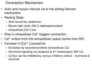
The Sliding Filament Mechanism • Actin and myosin slide past one another when the muscle contracts Evidence for this: • Sarcomere gets shorter • More overlap • Z-lines get closer together • I-band gets narrower • H-zone gets narrower
The role of ATP in muscle contraction The hydrolysis of ATP (adenosine triphosphate) provides the energy required for muscle contraction. 33kJmol-1energy + + inorganicphosphate ATP ADP Most muscle fibres store phosphocreatine, a chemical that phosphorylates ADP to ATP. This reaction maintains the muscle’s supply of ATP during vigorous exercise. + + ADP phosphocreatine ATP creatine
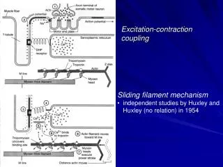
The sarcomere – structure to function Hansen and Huxley realized that the interlocking structure of the thick and thin filaments allows them to slide past one another. This reduces the length of the sarcomere. contraction At the same time the banding pattern of the sarcomere changes; light bands, formed by actin, shrink as the filaments become more interlocked. In 1954 Hansen and Huxley published their work explaining muscle contraction using their sliding filament theory.
The structure of myosin The myosin filament is formed from a number of myosin proteins wound together. Each ends in a myosin head, which contains an ATPase. myosin filament myosin head actin binding site ATP binding site ATPase head myosin neck
The structure of actin The actin filament is formed from a helix of actin sub-units. Each contains a binding site for the myosin heads. troponin myosin head binding site tropomyosin actin sub-unit Two other proteins are attached to the actin fibre: • tropomyosin is wound around the actin • troponin molecules are bound to tropomyosin and contain calcium ion binding sites.
Muscle Contraction – Sliding Filament Mechanism • Heads of myosin form cross-bridges with the actin filaments (attach to binding sites) • Myosin heads flex together and pull the actin along the myosin • They detach • Return to original angle and re-attach (uses ATP) • Repeats 100 times a second
Task • Using the play doh demonstrate muscle contraction • Four sections • Arrival of an action potential – Ca2+, troponin, tropomyosin, actin-myosin crossbridge • Movement of the actin filament – ATPase, ATP, power stroke • Breaking of the cross bridge – ATP, myosin head • Return to resting state – troponin, Ca2+, sarcoplasmic reticulum Use keywords – write down a flowchart explaining each stage
2. Anaerobic respiration in muscle cell sarcoplasm Produces lactate and can lead to fatigue/cramp. Maintaining ATP supply • Aerobic respiration in muscle cells mitochondria • Needs a supply of respiratory substrate and oxygen 3. Creatinine phosphate – another chemical present in muscle cells can donate its phosphate to recharge ADP back to ATP (supports a further 2-4 seconds
Plenary • Q10 p111
Learning Objectives Success Criteria • To understand the sliding filament model • Outline the role of ATP in muscle contraction (Grade E - D) • Outline how the supply of ATP is maintained in the muscle (Grade C –B) • Explain using diagrams the sliding filament model of muscular contraction (Grade B – A)
Sliding filament theory • When a nerve impulse arrives at a neuromuscular junction, calcium ions are released from the sarcoplasmic reticulum. • The calcium ions diffuse through the sarcoplasm. • This initiates the movement of the protein filaments as follows: • Calcium ions attach to the troponin molecules causing them to move. • As a result, the tropomyosin on the actin filament shifts position, exposing myosin binding sites on the actin filaments. • Myosin heads bind with myosin binding sites on the actin filament, forming cross bridges. • When the myosin head binds to the actin, ADP and Pi on the myosin head are released. • The myosin changes shape, causing the myosin head to nod forward. This movement results in the relative movement of the filaments. The attached actin moves over the myosin. • An ATP molecule binds to the myosin head. This causes the myosin head to detach. • An ATPase on the myosin head hydrolyses the ATP forming ADP and Pi. • This hydrolysis causes a change in the shape of the myosin head. It returns to its upright position. This enables the cycle to start again.