Download
1 / 55
571 likes | 1.51k Views
Learn about the components, structure, and functions of connective tissue cells and ground substance. Explore the origin, types, and characteristics of connective tissues.

E N D
ConnectiveTissue: cells and groundsubstance Dr. Anna L. Kiss Department of Anatomy, Histology and Embryology Semmelweis University Budapest 2017
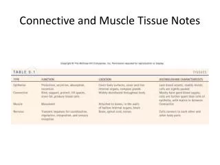
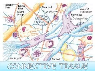
Connetivetissue Variety of tissueswithdifferentfunctionalproperitesbutwithcertaincommoncharacteristics Function: connectsorgans, fillsinspaces, storage, protection Structure: cells+extracellularmatrix Origin: mesoderm Fewercells, inloosearrangement; extracellularmatrix is thedominantcomponent Extracellularmatrix: - fibers:collagen elastic reticular - groundsubstance: proteoglycans, glycosaminoglycans, adhesionmolecules, inorganicsalts)
Connectivetissue Carola: Human Anatomy & Physiology
Connectivetissuecomponents CellsExtracellular matrix mobile fix fibers ground substance proteoglycans glycosaminoglycans adhesion glycoproteins fibrocyte reticular cell adipocyte melanocyte mesoblast macrophag mast cell plasma cell granulocyte (neutrophil, eosinophil) collagen elastic reticular fibrillin hyaluronic acid chondroitin sulphate heaparan sulphate keratan sulphate dermatan sulphate fibronektin laminin tenascin enactin trombospondin
Fix cells • fibrocyta (inact.), fibroblast (act.)– fiber (collagen+elastic) and matrix protein synthesis In adult: inactive fibrocytes, injury: growth factors: activation regeneration • reticular cells: - produce reticular fibers (spleen lymph nodes, red bone marrow) Collagen fibers
Fix cells • adipocytes – store lipids - ATP production - thermoregulation (especially brown fat tissue) brown adipocyte Röhlich Pál: Szövettan
Mobile cells • „immune cells” macrophag – phagocytosis, local defence, antigen-presentation granulocytes – phagocytosis lymphocytes– cellular and humoral immunity plasma cells – immunoglobulin (antibody) synthesis mast cell – ? allergy
Mobile cells Macrophages: 15-20µm • Defence: pathogen phagocytosis (FcR) lysosomal digestion vesicular transport to the cell surface: antigen presentation • „Cleaning”: phagocytosis of the dead cells • Secretion: growth factors(IL1, IL6) activation of the immune cells
Mobile cells Mast cells: 0.5-1µm Granules: heparin: blood clotting histamin: allergy Degranulation: regulated exocytosis IgE receptor-mediated signal transduction Lymphocytes, plasma cells: antibody synthesis Granulocytes: neutrophyl: phagocytosis eosinophyl: allergy
Extracellularmatrix • Proteoglycanes+adhesion molecules • filamentuos molecules: 3 dimensional network – gelatinuos material • is able to bind large amount of water – flexibility (turgor)
Proteoglycan GAG chains HA-binding region Core protein hyaluronic acid link protein
Proteoglycanes • Membrane-proteoglycanes: • Syndecan • CD44 • Trombomodulin • ECM-proteoglycanes: decorin biglican serglycine versican aggrecan
Fibers: collagenfiber • the most frequently present fibers • flexible, have remarkably high tensile strength • composed of collagen: many subtypes (I-XV), tissue specific distribution Synthesis: triple helix, composed of 3 α-chains procollagén In the extracellular matrix enzymatic hydrolisistropocollagén
Reticularfibers • fine, branching network • framework of the haemopoetic, and lympatic organs • special staining: silver impregnation • IV. típ. collagen • reticulum cells
Mesenchymal tissue Star-shapedmesenchymalcells and fibers: embryo, youngdevelopingtissue
Types of connectivetissue • loose connective tissue • few fibers, irregular structure • the most frequent type, • fills in spaces, connects organs with each other
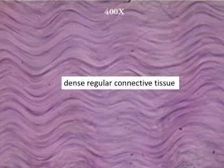
dense regular connective tissue many collagen fibers in regularly arranged bundles: tendons dense irregular connective tissue many collagen fibers, in irregularly arranged bundles
adipose tissue • adipocytes, a few reticular fibers • „Fat pad” • white and brown adipose tissue brownadiposetissue
Supporting tissue • Function: supports the body • Structure: cells + extracellular matrix • Origin: mesoderm • Types: cartilage bone
Cartilage CellsExtracellular matrix chondroblasts chondrocytes osteoblasts osteocytes osteoclasts groundsubstance fibers
Cells + extracellular matrix Cells: chondrocytes Extracellular matrix: fibers (collagen, elastic) + ground substance ground substance: proteoglycans glycoproteins: chondrocalcin, cartilage matrix protein (CMP), chondronectin fibronectin
Extracellular matrix • Proteoglycanes+adhesion molecules • filamentuos molecules: 3 dimensional network – gelatinuos material • is able to bind large amount of water – flexibility (turgor)
GAG: • Hyaluronicacid: glucuronicacid + N-acetilglucosamin • Chondroitinsulfate: N-acetyl-galactosamin • Keratansulfate: N-acetyl-glucosamin + sulfatedgalactose Proteoglycane: „core” protein + chondroitinsulfate and keratansulfate
Types of cartilage • Hyalin: functional unit: chondron: covers the bony surfaces of the joints • Elastic • Fibrous: intervertebral disc, temporomandibular joint
Chondron: chondrocytes basophylic capsule territorial matrix
General characteristics Mechanically resistent, hard, but relatively elastic and light skeletal structure. Highly vascularized living tissue, adaptable to changing environmental conditions, continuously being remodeled and renewed Components I. Cells (osteocytes, osteoblasts, osteoclasts, osteoprogenitor cells) II. Extracellular matrix (collagen fibers, proteoglycans, bone-specific proteins) + mineral salts: hydroxyapatite crystals
I. Cells in the bone tissue osteoprogenitor osteoblast osteocyte osteoclast
1. Osteocytes Bone cells with many thin processes (connected with similar processes of neighboring bone cells by gap junctions). Maintains the surrounding bone matrix by synthesizing or resorbing matrix constituents, if needed. Situated in small cavities of the matrix: lacunae, canaliculi.
2. Osteoblasts Cells synthesizing and secreting bone matrix components during development and restructuring of the bone. Sitting on the bone surface (often in a monolayer), they have a basophilic cytoplasm (rER!) and several processes penetrating into the newly formed matrix (osteoid). They are gradually embedded into the forming matrix and become osteocytes. At the end of osteogenesis, the remaining osteoblasts die with apoptosis or dedifferentiate into osteoprogenitor cells. Osteoblasts can induce the formation of osteoclasts on hormonal effects. osteoblast osteoid mineralized bone tissue
3. Osteoprogenitor cells Flattened cells with oval nuclei, situated on the peri- and endosteal surfaces and around blood vessels in the bone. Derivatives of mesenchymal stem cells and committed to bone formation. Capable of cell division and to form not only osteoblasts, but chondroblasts and fibroblasts as well. Endosteum, HE staining.
4. Osteoclasts Giant cells with the capacity of degrading the bone tissue. Multinucleated cell with an eosinophilic cytoplasm rich in mitochondria and lysosomes. They derive from blood monocyte-like cells which fuse and form osteoclasts on contact with osteoblasts (parathyroid hormone, RANK, signal transduction, gene activation, …) The cell surface facing the bone tissue has a ruffled border, consisting of many lamellar processes. Proton pumps of the membrane on this side pump H+ into the subcellular cavity, lysosomes are exocytosed releasing degrading enzymes, which leads to the degradation of both organic and inorganic components of the matrix. A ringlike peripheral zone of junctional structure (sealing zone) between the cell and the bone surface closes up the subcellular cavity.
II. Bone matrix 1. Collagen fibers.Main components (90%) of the organic matrix. Type I collagen. Parallelly arranged they often form 3-5 μm thick lamellae (in lamellar bone), but can be present as dense, woven structures (in woven bone). Collagen fibers are responsible for elastic resistence against bending and loading. Genetic defects of the collagen can lead to osteogenesis imperfecta (frequent fractures, deformities of the skeletal system). 2. Proteoglycans (PGs).They form a macromolecular network into which collagen fibers are embedded. PGs have shorter core proteins and fewer glysoaminoglycan (GAG) chains (mostly chondroitin 4,6 sulfate, keratansulfate) than in cartilage. 3. Bone-specific proteins.Osteocalcin and osteonectin (involved in mineralization), osteopontin (mediates the formation of the sealing zone in osteoclasts), bone sialoprotein (involved in attachment of osteoblasts to the matrix surface). 4. Inorganic material (mineral salts)makes 65% of the dry weight. Hydroxyapatite crystals: [3 Ca3(PO4)2 . Ca(OH)2], fine needle-like (40x2 nm) crystals bound to collagen fibrils with longitudinal orientation. Responsible for pressure resistence. Formation of the matrix:osteoblasts synthesize components of the organic matrix and regulate mineralization. Degradation of the matrix:osteoclasts degrade and dissolve both organic and inorganic constituents of the matrix.
Osteon Lamellae are arranged in concentric layers with layers of osteocytes between them. Canaliculi (containing osteocyte processes) form radial diffusion pathways for nutrients from the Havers canal up to the osteon periphery. Haversian (concentric) lamellae interstitial lamellae Haversian canal Osteon in cross section, Schmorl staining Osteons with polarized light
Periosteum Dense connective tissue covering on the bone. Outer layer:dense lamellar connective tissue,inner layer: in actively growing bone contains osteoprogenitor cells and osteoblasts, in inactive periosteum only few osteoprogenitor cells are present. No periosteum on articular surfaces. Blood vessels (nutrition of the bone tissue) and nerves (inflammation, damage causes strong pain!) Endosteum Thin connective tissue layer covering all internal surfaces (lining the marrow cavity, the trabecular surfaces and the canals inside the bone). Usually one layer thick with osteoprogenitor cells.
Haversian system in compact bone outer circumferential lamellae compact bone osteonal lamellae inner circumferential lamellae spongy bone Haversian canal with blood vessel Volkmann’s canal with blood vessel
Bone development 1.) Intramemebranous ossification: connective tissue 2.) Chondrogenic ossification: cartilage
Intramembranous ossification mesenchymal cells osteoblasts osteocytes
Intramembranous ossification At the surface of the osteoid, osteoblasts continue the appositional deposit of matrix, mainly type I collagen and groud substance proteins