Spinal Nerves
Spinal Nerves. Cervical plexus Brachial plexus. Objectives. Describe how spinal nerves are formed. Make a list of contributing roots to cervical plexus. Discuss the general arrangement. Describe the location of this plexus. Make a list of the out coming nerves..

Spinal Nerves
E N D
Presentation Transcript
Spinal Nerves Cervical plexus Brachial plexus
Objectives • Describe how spinal nerves are formed. • Make a list of contributing roots to cervical plexus. • Discuss the general arrangement. • Describe the location of this plexus. • Make a list of the out coming nerves.. • Point out the point where the major cutaneous nerves emerge. • Describe the brachial plexus • Make a list of contributing spinal nerves. • Discuss the general arrangement of this plexus. • Locate the plexus in the axilla and note important relations to blood vessels. • Make a list of the terminal main branches of brachial plexus.
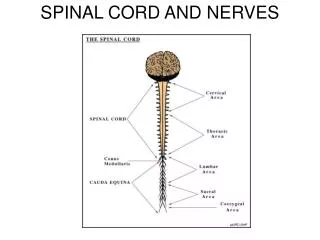
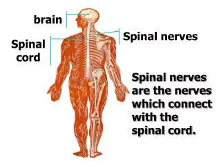
Spinal Nerves • The spinal cord is functionally segmented • Each segment gives of a pair of spinal nerves (31) • 8 cervical, 12 thoracic, 5 lumbar, 5 sacral & one coccygeal • Each nerve has a dorsal (sensory) root & a ventral motor root • The dorsal has a ganglion containing pseudounipolar neurons
Each spinal nerve arises as rootlets which then combine to form dorsal (posterior) & ventral (anterior) roots. • Two roots merge laterally and form the spinal nerve. • Dorsal (posterior) root has a ganglion (dorsal root/sensory ganglion) that contains the cell bodies of the sensory neurons • Each spinal nerve then divides into a smaller dorsal and a larger ventral ramus
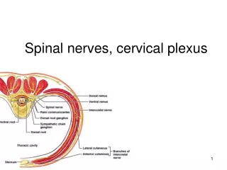
Spinal nerve Typical spinal nerve divides into: • 1- Dorsal ramus which supplies structures on the back • 2- White ramus communicans to sympathetic trunk and Grey ramus communicans from the sympathetic trunk • 3- Collateral branch • 4- Ventral ramus • 5- Lateral branch • 6- Anterior branch
Main Branches of the spinal nerve • Dorsal Ramus: innervate deep muscles of the trunk responsible for movements of the vertebral column and skin near the midline of the back. • Ventral Ramus: what they innervate depends upon which part of the spinal cord is considered. • Thoracic region: form intercostal nerves that innervate the intercostal muscles and the skin over the thorax • Remaining spinal nerve ventral rami (roots of the plexus): form five plexuses (intermingling of nerves). • Ventral rami of C1-C4= cervical plexus • Ventral rami of C5-T1= brachial plexus • Ventral rami of L1-L4= lumbar plexus • Ventral rami of L4-S4= sacral plexus • Ventral rami of S4 and S5= coccygeal plexus
Communicating Rami: communicate with sympathetic chain of ganglia.
Dermatomes • Dermatome is: A specific segment of skin supplied by a single spinal nerve. • All spinal nerves except for C1 innervate a segment of skin, and so each of these nerves is associated with a dermatome. • The skin of the body may be divided into sensory segments that collectively make up a dermatome map.
Dermatomes • Cutaneous areas supplied by adjacent spinal nerves overlap. There is therefore littleor nosensory loss after interruption of a single spinal nerve or dorsal root
Cervical Plexus • Is made of the anterior rami of C1-4 • Ansacervicalis: is a nerve loop made of superior root C1-2 and inferior root C2-3 • Ansacervicalis and anterior ramus of C1 supply infrahyoid muscle • Phrenic nerve arises from C3-5
Cervical Plexus Sensory branches of cervical plexus include: • Lesser occipital nerve • Great auricular nerve • Transverse cervical nerve • Supraclavicular nerves
Brachial Plexus • It is formed by the ventral rami of C5-T1 • C5-6 form upper trunk • C7 continues as middle trunk • C8-T1 form the lower trunk • Each trunk divides into anterior and posterior divisions
FORMATION OF BRACHIAL PLEXUS • It is formed in theposterior triangleof the neck. • Divisions: • The plexus is divided into : • Roots • Trunks • Divisions • Cords • Terminal branches
TRUNKS • Upper trunk • Union of the rootsofC5 & 6 • Middle trunk • Continuation of theroot ofC7 • Lower trunk • Union of the rootsofC8 & T1
DIVISIONS & CORD • Each trunk divides into anterior and posterior division • Posterior cord: • From the three posterior divisions • Lateral cord: • From the anteriordivisions of the upperand middle cords
CORDS & BRANCHES • Medial cord • It is the continuation of the anterior division of the lower trunk • Branches • All three cords will give branches, those will supply their respective regions
BRANCHES • (A) From Roots: 1. C5:Nerve torhomboids (dorsal scapular nerve). 2. C5,6 &7:Longthoracic nerve • (B) From Trunk (upper trunk): • Nerve to subclavius • Suprascapularnerve (supplies supraspinatus & infraspinatus)
(C)BRANCHES From Cords Lateral Cord (2LM) .Lateral pectoral n .Lateral root to median n .Musculocutaneous n C5 C6 C7 C8 T1 Medial cord (4MU) .Medial pectoral n. .Medial root to median n. .Medial cutaneous n of arm. .Medial cutaneous n of forearm. .Ulnar n. Posterior Cord (ULTRA) .Upper subscapular n .Lower subscapular n .Thoracodorsal n .Radial n .Axillary n