Structure and Function of Macromolecules in Biology
430 likes | 460 Views
Learn about the four classes of macromolecules - Carbohydrates, Lipids, Proteins, and Nucleic Acids. Understand polymerization and hydrolysis reactions, as well as the structural variations of macromolecules. Explore the functions and characteristics of carbohydrates and lipids in living organisms.

Structure and Function of Macromolecules in Biology
E N D
Presentation Transcript
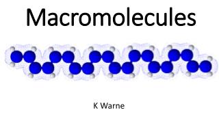
Chapter 5: Structure & Function of Macromolecules • Most macromolecules are polymers. which is not? • Polymer: large molecule consisting of many identical or similar subunits connected together in repeating fashion • Monomer: the building block molecule of a polymer
Macromolecule: a large organic polymer • There are four classes of macromolecules: • Carbohydrates • Lipids • Proteins • Nucleic acids
macromolecules are synthesized by polymerization reactions. A type of synthesis reaction Also called a condensation rxn ordehydration rxn
Condensation reactionsreactions during which monomers are covalently linked, producing a net removal of one water molecule per covalent linkage. One monomer loses a hydroxyl (-OH) and the other loses a hydrogen (-H) REQUIRES ENERGY!!! Requires biological catalysts (enzymes)
How are macromolecules broken? • Hydrolysis is a reaction process that breaks covalent bonds between monomers by the addition of water. Also called a decomposition reaction. A hydrogen from the water bonds to one monomer, and the hydroxyl bonds to the other monomer. Digestive enzymes are hydrolytic
Structural variation only40-50 common monomers are used to form biological macromolecules.
I. Carbohydrates – organic molecules made of sugars and their polymers Consists of the elements C, H, O The monomers =monosaccharides. (C H2 O) Are classified based on the number of simple sugars mono- , di- , poly-
A. Monosaccharides– Simple sugars in which C, H, and O occur in the ratio of: CH2O (glucose – C6H12O6) Are the major nutrients for cells [Glucose ) • Can be produced by photosynthesis
Characteristics of a sugar: 1. A hydroxyl is attached to each carbon except one, which is double bonded to an oxygen (carbonyl) • 2. Size of the carbon skeleton varies from 3 to 7 carbons. The most common monosaccharides are: • Classification # of carbonsexample • Triose 3 Glyceraldehyde • Pentose 5 Ribose • Hexose 6 Glucose
3. Spatial arrangment around asymmetric carbons may vary. For example, glucose and galactose 4. In aqueous solutions, many monosaccharides from rings.
B. Disaccharides – a double sugar that consists of two monosaccharides joined by a glycosidic linkage. A glycosidic linkage is a covalent bond(5.5] • Common examples of disaccharides: Maltose – Glucose + Glucose (brewing) Lactose – Glucose + Galactose (present in milk) Sucrose – Glucose + Fructose (table sugar)
C. Polysaccharides – polymers of a few hundred or thousand monosaccharides Formed by condensation reactions Have two important biological functions. Energy storage (starch and glycogen) Structural support (cellulose and chitin)
Storage polysaccharides: Cells hydrolyze storage polysaccharides into sugars as needed. Starch is a storage polysaccharide in plants. Major sources are potato and grains (wheat, corn, rice, etc.) Glycogen is a storage polysaccharide in animals. stored in the muscle and liver
Structural polysaccharides: Cellulose is a major component of plant cell walls. - reinforces plant cell walls using H-bonds. -can’t be digested by most organisms Chitin forms exoskeletons of arthropods and the cell walls of some fungi.
L I P I D S insoluble in H2O, but will dissolve in nonpolar solvents.fats, phospholipids, and steroidsA. Fats – macromolecules that consist of:- Glycerol, a three-carbon alcohol -Fatty acids, a carboxylic acid
A fatty acid- carboxyl group at one end and an attached hydrocarbon chain (“tail”) [Fig. 5.10] The carboxyl functional group (“head”) has acidic properties. The hydrocarbon chain has a long carbon skeleton usually with an even number of carbon atoms (most 16 – 18)The nonpolar C-H bonds make the chain hydrophobic and water insoluble.
condensation reactions link glycerol to fatty acids by an ester linkage. An ester linkage is a bond between a hydroxyl group and a carboxyl group.Each of glycerol’s three hydroxyl groups can bond to a fatty acid by an ester linkage producing a fat.
T Y P E S of FATS A)Triacylglycerolis a fat composed of three fatty acids bonded to one glycerol by ester linkages (triglyceride)Fatty acids may vary in the number and location of carbon-to-carbon double bonds:
SATURATED FAT No double bonds between carbons in FA tail Carbon skeleton of FA is“saturated” w/ hydrogens)Usually solid at room temp. Most animal fats- Bacon grease, lard, butter
Unsaturated fats Tail kinks at each C=C, so molecules do not pack close enough to solidify at room temp. Usually liquid at room temp. Most plant fats- Corn, peanut, and olive oil Are there any unsaturated fats that are solid at room temp?Hydrogenated or trans-fats
Triglyceride FUNCTIONS Energy storage(1g of fat stores twice as much energy as 1g of polysaccharide) Animals store moreenergy with lessweight than plants which use starch. Oxygen is heavy!! Cushions vital organsin mammals (e.g. kidney)Insulates against heat loss
B. Phospholipids– compounds with molecular building blocks of glycerol, two fatty acids, and a phosphate group (usually w/ a polar molecule attached) [Fig 5.12]third carbon of glycerol is joined to a negatively charged phosphate group.Are amphipathic (have hydrophobic and hydrophilic ends).
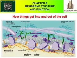
Phospholipids are….. • The major constituents of cell membranes. form a bilayer held together by hydrophobic interactions among the hydrocarbon tails. If placed in water, a “micelle” can form. [Fig 5.13
C. Steroids– lipids which have four fused carbon rings with various functional groups. example: Cholesterol 1) precursor to sex hormones, 2) component in animal cell membranes 3) contributes to atherosclerosis
PROTEIN III. Protein– polymer (chain) of amino acidsFunctions Structural supportStorage(of amino acids)Transport (e.g. hemoglobin)Signaling (chemical messengers)Movement(contractile proteins)Cellular response (receptor proteins) Defense (antibodies)Catalysis of biochemical reactions(enzymes)
What’s an amino acid? consist of a carbon (alpha carbon) bonded to: a) Hydrogen atomb) Carboxyl groupc) Amino groupd) R group (a side chain that makes each amino acid unique)
The twenty amino acids can be grouped by properties of the side chain (Fig 5.15) Nonpolar side groups (hydrophobic). Polar side groups (hydrophilic) - uncharged polar- charged polar
How are polypeptide chains formed? amino acid monomers are covalently linked by peptide bonds. [Fig 5.16] Formed by a condensation reaction that links the carboxyl group of one amino acid to the amino group of another.How are polypeptide chains broken?
A protein’s function depends upon its unique structure. Primary structureSecondary structureTertiary structureQuaternary structureThese four levels of protein structure are responsible for the correlation between form and function in proteins.
Primary Structure– unique sequence of amino acids in a proteinDetermined by genes Slight change can affect a protein’s conformation and function (e.g. sickle-cell hemoglobin; Fig 5.19) Can be sequenced in a laboratory; Fredrick Sanger (insulin sequence in late 1940’s, early 1950’s)
Secondary Structure– regular, repeated coiling and folding of a protein’s polypeptide backbone. Contributes to protein’s overall conformation Stabilized by hydrogen bonds between peptide linkages in the protein’s backbone (carbonyl & amino groups).
Two major types of secondary structure: Alpha Helix – secondary structure of a polypeptide that is a helical coil stabilized by hydrogen bondingBeta sheet – secondary protein structure which is a sheet of antiparallel chains folded like an accordian. Parallel regions are held together by hydrogen bonds
Tertiary Structure– irregular foldings of a protein due to bonding between side chains (R groups)1-H-bonding between polar side chains2-Ionicbonds between charged side groups3-Hydrophobic interactions between nonpolar side chains in protein’s interior4-Covalent linkages5-Disulfide bridges between cystine monomers
Quaternary Structure – results from the interaction amongseveral polypeptide(subunits) in a single proteinExamples: collagen, hemoglobin
If a protein’s environment is altered, it may become denatured. Denaturation is a process that alters a protein’s native conformation and biological activity. Proteins can be denatured by:1-Chemical agents that disrupt H-bonds, ionic bonds, and disulfide bonds2-Excessive heat3-Transfer to an organic solvent. The hydrophobic chains that were originally inside, turn outward
Predicting 3-dimensional shape of proteins is difficult!! Chaperone proteins have been discovered that temporarily brace a folding protein
Nucleic Acids – polymer of nucleotides liked together by condensation reactions I.Deoxyribonucleic Acid (DNA)-Contains coded informationthat programs all cell activity(genes)-Contains directions for its own replication-Is copied and passed from one generation of cell to another-Found in the nucleus of eukaryotes
II. Ribonucleic Acid (RNA)Functions in the actual synthesis of proteins coded for by DNA-Sites of protein synthesis are on ribosomes in the cytoplasm-Messenger RNA (mRNA) carries encoded genetic message from the nucleus to the cytoplasm
A nucleotide is a building block molecule of nucleic acid, made up of 1) a five carbon sugar covalently bonded to 2) a phosphate group and 3) a nitrogenous base The 5-carbon sugar can be either ribose or deoxyribose.
There are two families of nitrogenous bases:PyrimidineCytosine (C) Thymine (T) – found only in DNAUracil (U) – found only in RNAPurineAdenine (A)Guanine (G)
A nucleic acid is a polymer of nucleotides joined by phosphodiester linkages between the phosphate of one nucleotide and the sugar of the next. Each gene on DNA contains a uniquelinear sequence of nitrogenous bases which ultimately codes for a uniquelinear sequence of amino acids in a protein.
In 1953, James Watson and Francis Crick proposed the double helix as the three dimensional structure of DNAthe sugar-phosphate backbones are on the outside of the helixnitrogenous bases are paired in the interior of the helix and are held together by hydrogen bonds.A always pairs with T; G always pairs with CThe two strands of DNA are complimentary, thus can serve as templates to make new complimentary stands.