Download
1 / 14
140 likes | 1.04k Views
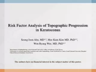
Rotating Scheimpflug Topographic Parameters Important in Distinguishing Keratoconus Suspect from normal eyes. Clayton Falknor, MD, Orkun Muftuoglu, MD, R. Wayne Bowman , MD, Steven Verity, MD, James P. McCulley, MD

E N D
Rotating Scheimpflug Topographic Parameters Important in Distinguishing Keratoconus Suspect from normal eyes Clayton Falknor, MD, Orkun Muftuoglu, MD, R. Wayne Bowman, MD, Steven Verity, MD, James P. McCulley, MD Some of the authors have received consultant reimbursement from Alcon Labs, Inc. None of the authors have financial interest in the subject matter of this poster.
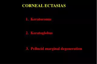
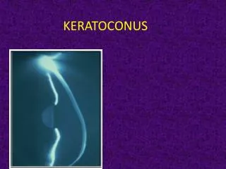
Iatrogenic corneal ectasia • Complication of corneal refractive surgery • Progressive corneal steepening, myopia, astigmatism, loss of BCVA • Pre-operative risk factors • Thin residual stromal bed after LASIK • Forme fruste keratoconus or keratoconus • Keratoconus is characterized by: • Non-inflammatory, progressive corneal thinning and bulging • Irregular astigmatism and myopia • Potentially severe corneal scarring • Keratoconus is identified by: • Examination findings (Fleisher ring, Vogt striae, subepithelial fibrosis, stromal thinning, scissoring of retinoscopic reflex) • Central or paracentral steepening on topography
Subclinical keratoconus • Traditional method to identify subclinical keratoconus is Placido disk-based corneal topography • Reflection-based system • Measures slopes of anterior corneal surface only • Axial curvature method subject to misalignment of corneal apex and corneal sighting point • Contribution of posterior corneal surface important • Projection-based systems • Orbscan (Bausch and Lomb, Salt Lake City, Utah, USA) • Pentacam (Oculus Optikgeraete GmbH, Germany)
Keratoconus suspects • Topographic designation • No evidence of keratoconus on examination • Multiple methods to define • Modified Rabinowitz/McDonnell method (central K steeper than 47.2D, I-S >1.4D) • Maeda/Klyce KPI index • Smolek/Klyce KSI index (based on >10 indices and neural network) • KISA% (Rabinowitz)
Pentacam Comprehensive Eye Scanner • Rotating Scheimpflug camera • Monochromatic slit light source rotates with camera • 25-50 slit images per acquisition • Eye movement monitoring by 2nd camera • Less than 0.6mm decentration • Rotates 180º in 2 seconds • All images include central cornea • Corneal elevation data independent of visual axis and corneal apex http://www.oculus.de/chi/downloads/dyn/sonstige/sonstige/pentacam_aao_2006.pdf
Purpose • Evaluate Pentacam parameters important in distinguishing keratoconus suspects from normal • Pentacam parameters to detect keratoconus • Pachymetry • Progression index of corneal thinning • Corneal volume within fixed diameter • Keratometry readings and axis • AC volume, depth and angle • Posterior elevation over best-fit sphere (option of toric ellipsoid) • Zernike HOA of anterior and posterior surfaces • Corneal variance indices • ISV (index of surface variance) • IVA (index of vertical asymmetry) • IHA (index of height asymmetry) • IHD (index of height decentration) • Rmin (radius minimum) • KI (keratoconus index) • CKI (center keratoconus index)
Patients • Controls (normals presenting for keratorefractive surgery) • 72 eyes of 41 patients • Inclusion: underwent pre-operative screening for keratorefractive Sx, normal corneal exam, available topography maps • Exclusion: prior ocular surgery or trauma, ocular disease likely to affect corneal HOA’s • Keratoconus suspects • 15 eyes from 10 patients • Selected from normals with keratoconus suspect indication by Smolek/Klyce KSI on topography. • Keratoconus (diagnosed clinically with topography support) • 108 eyes of 54 patients (34 men, 20 women) • Inclusion: distorted keratometry mires, abnormal retinoscopic reflex, Vogt’s striae, Fleischer’s ring, corneal scarring, available topography maps • Exclusion: prior corneal surgery, extensive corneal scarring
Posterior corneal elevation • Mean posterior elevation • Keratoconus 98.7 ± 46.3 µm • Keratoconus suspects 16.9 ± 6.1 µm • Controls 8.6 ± 3.8 µm
Summary of results • Maximum posterior corneal elevation • For keratoconus • Cut-off of 35 µm, sensitivity 93% and specificity 95% • For keratoconus suspect • Cut-off 15 µm, sensitivity 67% and specificity 91% • Cut-off 10 µm, sensitivity 93% and specificity 61% • Progression index average and maximum all significantly different in keratoconus suspects vs. controls • Other significant parameters: • Corneal variance parameters : KI, IHD, Rmin • Pachymetry at pupil center and thinnest • Keratometry (flat and steep) • AC depth
Zernike analysis • Both anterior and posterior elevation data decomposed into Zernike higher-order aberration polynomials • Real differences between keratoconus and controls within the third through sixth orders • Anterior surface: vertical coma, trefoil • Posterior surface: vertical coma, spherical aberration, and fifth-order vertical coma
Pentacam for keratoconus • Pentacam is useful for identifying keratoconus suspects • Corneal variance parameters are set too high to capture keratoconus suspects, but specificity is excellent for confirmation purposes • Best Posterior elevation cut-off between 10 and 15 µm. • Important parameters include pachymetry, progression of corneal thinning, keratometry, corneal variance indices, and Zernike HOA’s (especially vertical coma)