Download
1 / 23
330 likes | 2.28k Views
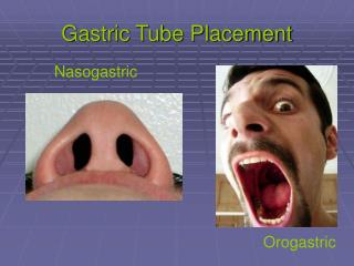
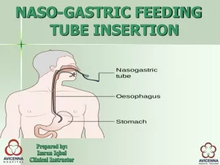
Naso-gastric tube insertion. Mem Van Beek Clinical Educator Bradford Teaching Hospitals. AIM. To enable the student to understand the principles of safe NG tube use. Objectives. By the end of this session students should be able to: State: Types of NG tubes & their uses

E N D
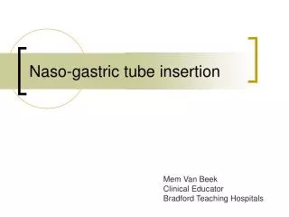
Naso-gastric tube insertion Mem Van Beek Clinical Educator Bradford Teaching Hospitals
AIM • To enable the student to understand the principles of safe NG tube use.
Objectives • By the end of this session students should be able to: • State: • Types of NG tubes & their uses • Indications for insertion • Complications • Legal aspect around NG tube insertion • Insert a naso-gastric tube safely and competently
Types of NG tubes Ryle’s tube for gastric drainage Fine –bore feeding tube Feeding Draining
INDICATIONS • FINE BORE NG TUBE • Short term enteral feeding (4-6 weeks) • Malnutrition • Head & neck surgery • Ca Head & neck / oesophagus • Inadequate intake • Oral cavity fistulae • To prolong & sustain life
INDICATIONS cont • RYLE NG TUBE • To drain gastric contents • Abdominal distension • Unconscious pt • Major surgery • Intestinal obstruction • To stop vomiting & prevent aspiration
Contraindications • Head injury – basilar skull # • Rhinorrhea –CSF • Obstructing oesophageal ca • Epistaxis • Feeding above an obstruction • Recent gastro oesophageal anastomosis • Hx of nasal or sinus surgery • occlusions
Cautions • Neck & buccal flap repair • Laryngectomy • Oesophageal ca • Head & neck surgery • Uncooperative pts
Complications of NG feeding • Aspiration • due to feed regurgitation • or incorrect tube placement • Nausea & vomiting • due to rapid feeding • poor gastric emptying • Diarrhoea • Type of feed ie Jevity • Gut infection
Complications cont • Constipation • inadequate fluid intake • immobility • use of opiates • Blocked tube • inadequate or no flushing of tube • administering meds via tube • Unstable BMs • ↑BMs esp with high carb feed • ↓BMs esp if feed is stopped quickly or interupted
Complications cont • Deranged electrolytes- re feeding syndrome • Fluid overload • Intestinal obstruction • Dislodged tube • Weight loss/ gain • Due to feed imbalances – poor regime • Excoriation of skin around tube
Risks associated with NG tubes • Pneumothorax • Coiling of tube in the throat • Parotiditis • Retropharyngeal Abscess • Sinusitis • Acid reflux • Aspiration pneumonitis • Severe sepsis (the most serious risk)
Legal Aspect • 2005 NPSA – 11 deaths due to misplaced NG feeding tubes • Correct & clear documentation • National & Local guidelines
Measuring length of feeding tube From bridge of nose to ear lobe to bottom of xiphisternum
Equipment required • Tray • Fine bore with introducer / Ryle’s tube • Receiver • Sterile water • Glass of water • 20ml syringe • Tape (hypoallergenic) • Lubricating jelly • Indicator strips ( pH fix, 0-6, Fisher scientific)
Procedure • Clinically clean procedure • Wash hands • Introduce self • ID patient • Gain informed consent • Arrange a signal of communication • Pt to sit in high Fowler’s position • Prepare equipment • Measure tube (as previously stated) & mark with tape.
Procedure • Lubricate tube • Check for nostril patency • Insert the rounded end of tube into the clearer nostril & slide it backwards & inwards along the floor of the nose to the nasopharynx. • When tube reaches nasopharynx (back of throat), ask pt to sip & swallow some water using a straw. • Advance the tube through the pharynx (as pt continues to swallow) till the predetermined mark has been reached • If at any point pt shows signs of distress/ cyanosis – remove tube.
Procedure • Secure the tube to nostril & cheek with tape • Check the position of the tube to confirm that it is in the stomach by • Check pH • Do X-ray of chest & upper abdomen • NO OTHER METHODS ARE ACCEPTED (NPSA 2005) • If position is correct; • Mark the tube at the exit site & record the tube length in the notes • remove guide wire from fine-bore tube & start feeding per regime • Connect drainage bag to Ryle’s tube for free drainage or spigot for prn aspiration.
Checking pH • Flush the NG tube with 20ml of air – to clear any substance already in tube • Aspirate 2ml of stomach content and test on pH strip. (blue litmus paper should not be used) • pH should be ≤5.5 (acidic) • If checking pH in tube already in place, wait 1hour after feed or medication as these can affect pH reading. • If pH of >5.5 is obtained – & pt is asymptomatic send for X-ray
REMEMBER • DO NOT use the ‘whoosh’ test • DO NOT use blue litmus paper • DO NOT use absence of respiratory distress • DO NOT monitor bubbling at end of tube • DO NOT use appearance of fluid aspirate NPSA 2005
Document • Date • Time • Type of tube inserted • Reason • Length inserted & how it is marked • pH of aspirate • Nursing instructions