Download
1 / 12
120 likes | 267 Views
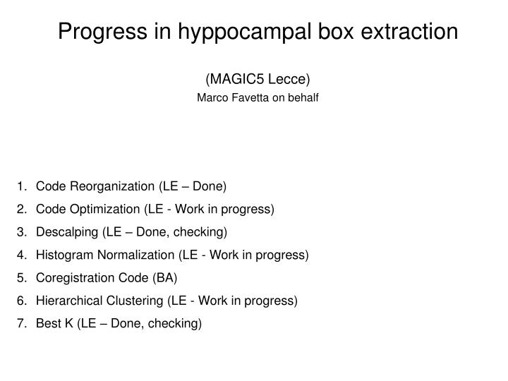
Progress in hyppocampal box extraction. (MAGIC5 Lecce) Marco Favetta on behalf. Code Reorganization (LE – Done) Code Optimization (LE - Work in progress) Descalping (LE – Done, checking) Histogram Normalization (LE - Work in progress) Coregistration Code (BA)

E N D
Progress in hyppocampal box extraction (MAGIC5 Lecce) Marco Favetta on behalf Code Reorganization (LE – Done) Code Optimization (LE - Work in progress) Descalping (LE – Done, checking) Histogram Normalization (LE - Work in progress) Coregistration Code (BA) Hierarchical Clustering (LE - Work in progress) Best K (LE – Done, checking)
Old code • Head Normalization (Pipeline module) • Exhaustive box extraction by ITK filter • Clustering by k-means • Non exhaustive box extraction
Code Reorganization BoxExtraction (main program) The code was reorganized as follows: Read config file Head normalization Exhaustivebox extraction Box coregistration(distance minimization) kmin…kmax K-means clustering Non-exhaustivebox extraction kmin…kmax Best k! Distance-based comparison kmin…kmax
Developments • Best K Find the smallest/best value of K according to the average distance of “k-means boxes” from “exhaustive boxes” • New coregistration filter, step by step (BA) Comparing different kinds of filters with the ITK filter. We think that using a step by step system we can reduce the errors due to possible rotation of the boxes.
Developments • Hierarchical clustering it does not require a priori choose of the number of clusters (k) • Descalping Significant improvement of normalization (Not only Head Normalization but Head + Brain normalization).
Hierarchical clustering Test on 49 boxes distances = {'euclidean'; 'seuclidean'; 'cityblock'; 'minkowski'; 'cosine'; 'correlation'; 'spearman'; 'hamming'; 'jaccard'; 'chebychev'}; methods = {'single'; 'complete'; 'average'; 'weighted'; 'centroid'; 'median'; 'ward'}; C = COPHENET(Z,Y) cophenetic correlation coefficient for the hierarchical cluster tree represented by Z. Z is the output of the function LINKAGE. Y contains the distances or dissimilarities used to construct Z, as output by the function PDIST. The cophenetic correlation for a cluster tree is defined as the linear correlation coefficient between the cophenetic distances obtained from the tree, and the original distances (or dissimilarities) used to construct the tree. Thus, it is a measure of how faithfully the tree represents the dissimilarities among observations. % 0.931100 jaccard average % 0.909639 jaccard single % 0.908437 hamming average % 0.897070 correlation average % 0.892561 spearman average …
Descalping Now Exhaustivebox extraction Future Descalping Hist. Normalization Exhaustivebox extraction
Brain extraction (descalping) • In: • K. Boesen, K. Rehm, K. Schaper, S. Stoltzner, R. Woods, D. Rottenberg, • Quantitative Comparison of Four Brain Extraction Algorithms, 9th International Conference on Functional Mapping of the Human Brain, • June 19- 22, 2003, New York, NY. Available on CD-Rom in NeuroImage, Vol. 19, No. 2. (http://www.neurovia.umn.edu/home/kelly/KB_HBM2003.pdf) • four brain extraction algorithms (BEA) are evaluated (web site links point to the latest versions): • Statistical Parametric Mapping (SPM), v. 2b (http://www.fil.ion.ucl.ac.uk/spm/software/spm8/) • Brain Extraction Tool (BET), v. 1.2 (http://www.fmrib.ox.ac.uk/analysis/research/bet/) • Minneapolis Consensus Strip (McStrip) (http://www.neurovia.umn.edu/incweb/) • Brain Surface Extractor (BSE), v. 2.99.8 (http://users.loni.ucla.edu/~shattuck/brainsuite/cortical-surface-extraction/skull-stripping-with-the-brain-surface-extractor-bse/)
Brain extraction (descalping) execution time is measured (no info on the used machine) and quality comparison with manual segmentation is performed: All of the images we have worked on till now (182 nifti files) were processed As our aim is brain coregistration, it is possible that small mistakes will not affect the final result: this is currently undergoing a full check According to this table, we chose BET (current version 2.1, a part of the FSL Software Library, http://www.fmrib.ox.ac.uk/fsl/) because very fast, even if the misclassified tissue % can be quite high. How many images suffer from tissue misclassification? How severe are these errors? How much will misclassification influence the brain coregistration stage?
Descalping We are testing these softwares: MRI Brain Segmentation (MATLAB) BET - Brain Extraction Tool
m1 p1 p2 m2 Histogram standardization MRI intensities do not have a fixed meaning, not even within the same protocol for the same body region obtained on the same scanner for the same patient. Simplest solution: transform image histograms by landmark matching, so that similar intensities will have similar tissue meaning Image histograms after descalping Images 1° group (1…133) • Determine location of landmarki (example: mode, median, various percentiles). • Map intensity of interest to standard scale for each volume image linearly and determine the location ’s of i on standard scale. Work in progress Images 2° group (134…182) L. G. Nyúl, J. K. Udupa, X. Zhang, New Variants of a Method of MRI Scale Standardization, IEEE TRANSACTIONS ON MED. IMAG., VOL. 19, NO. 2, FEBRUARY 2000 “standardization should be performed only after inhomogeneity correction, since the latter step can introduce its own intensity nonstandardness” (Kraft K, Fatouros P, Clarke G, Kishore P. An MRI phantom material for quantitative relaxometry. Magn Reson Med 1987;555–562.)