Download
1 / 21
230 likes | 548 Views
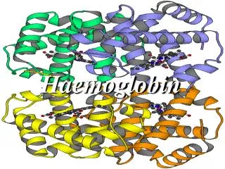
This overview delves into the essential role of hemoglobin (Hb) in red blood cells, primarily its function in oxygen (O2) and carbon dioxide (CO2) transport. Each red blood cell contains approximately 640 million Hb molecules, synthesized from globin and heme. The synthesis occurs at different cellular sites, with globin produced in polyribosomes and heme in mitochondria. The interaction of Hb with O2 is characterized by a sigmoid dissociation curve, influenced by factors like pH and 2,3-DPG. Understanding these mechanisms reveals the importance of Hb structure in maintaining oxygen delivery to tissues.

E N D
Introduction • The main function of red blood cell • Transfer of O2 from lungs to tissue • Transfer of CO2 from tissue to lungs • To accomplish this function red cells has haemoglobin (Hb) • Each red cell has 640 million molecules of Hb
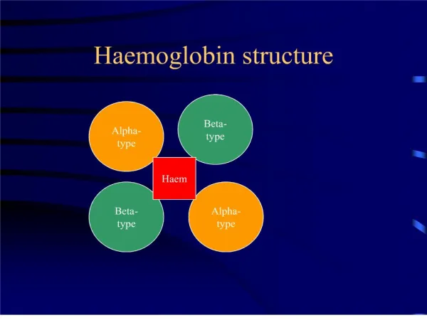
Introduction • Haemoglobin (Hb), protein constituting 1/3 of the red blood cells • Synthesis begins in proerythroblast • 65% at erythroblast stage • 35% at reticulocyte stage • Two parts • Haem • Globin
Synthesis of Haemoglobin (Hb) • Haem & globin produced at two different sites in the cells • Haem in mitochondria • Globin in polyribosomes • Well synchronized
Synthesis of Haem • Protoporphyrin ring with an iron atom in centre • The main site is mitochondria as it contains ALAS • Mature red cell does not contain mitochondria
Synthesis of globin • Various types of globin combines with haem to from different haemoglobin • Eight functional globin chains, arranged in two clusters the • b- cluster (b, g, d and e globin genes) on the short arm of chromosome 11 • a- cluster (a and z globin genes) on the short arm of chromosome 16
Synthesis of globin Globin synthesis, starts at 3rd week of gestation • Embryonic Haemoglobin Gower I ( z2e2) Haemoglobin Portland ( z2g2) Haemoglobin Gower II (a2e2) • Fetal : HbF (a2g2), HbA (a2b2) • Adult : HbA, HbA2 ( a2d2), HbF.
Functions of Haemoglobin • Oxygen delivery to the tissues • Reaction of Hb & oxygen • Oxygenation not oxidation • One Hb can bind to four O2 molecules • Less than .01 sec required for oxygenation • b chain move closer when oxygenated • When oxygenated 2,3-DPG is pushed out • b chains are pulled apart when O2 is unloaded, permitting entry of 2,3-DPG resulting in lower affinity of O2
Oxygen-haemoglobin dissociation curve • O2 carrying capacity of Hb at different Po2 • Sigmoid shape • Binding of one molecule facilitate the second molecule binding • P 50 (partial pressure of O2 at which Hb is half saturated with O2) 26.6mmHg
Hb-oxygen dissociation curve • The normal position of curve depends on • Concentration of 2,3-DPG • H+ ion concentration (pH) • CO2 in red blood cells • Structure of Hb
Hb-oxygen dissociation curve • Right shift (easy oxygen delivery) • High 2,3-DPG • High H+ • High CO2 • HbS • Left shift (give up oxygen less readily) • Low 2,3-DPG • HbF
Summary • Normal structure including the proportion of globin chains are necessary for the normal function of haemoglobin • Reduced haemoglobin in the red blood cells due to any abnormality of any of its constituents result into a clinical situation called anaemia • Metabolic & other abnormalities result into abnormal oxygen supply to the tissue